
| Sang-Bum Kim | 5 Articles |
Purpose
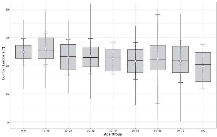
To evaluate the association between lumbar lordosis and age using an AI-based automated measurement model applied to a large dataset of standing lateral spinal radiographs. Materials and Methods This retrospective study analyzed 904 high-quality radiographs selected from 2,397 images acquired between 2019 and 2021. Lumbar lordosis was defined as the angle between the superior endplates of L1 and S1 and automatically measured using a validated deep learning model. Subjects were categorized into nine age groups. One-way ANOVA compared lumbar lordosis across age groups, and Pearson correlation assessed the relationship between age and lumbar lordosis. Results Lumbar lordosis ranged from 0° to 84° (mean 45.9°±13.4°). The highest mean value was in the 10–19-year group (52.1°), and the lowest in the ≥80-year group (39.6°). Minimum values decreased to 0° in individuals aged ≥60 years. No significant differences were found across age groups (p=0.561). A weak but significant negative correlation was observed between age and lumbar lordosis (r=–0.247, p<0.0001). Conclusions AI-based automated measurement enabled efficient large-scale analysis and revealed a wide distribution of lumbar lordosis with a gradual age-related decline. These findings highlight the value of AI in spinal alignment assessment.
Purpose
The current study aims to report the results of analyzed factors that ultimately undergo surgical treatment after selective nerve root block in patients with spinal structural pathology that cause lower back pain and radiating pain in the lower extremities. Material and methods: A retrospective study was performed on 537 patients diagnosed with spinal canal stenosis or disc herniation among patients who underwent selective nerve root block at our hospital for five years from May 2015 to December 2017. The patients were divided into Group A (patients with an only selective spinal nerve root, n=99) and Group B (patients with surgical treatment, n=20). We evaluated the primary demographic factors, including age, sex, onset, symptom duration, diabetes mellitus, hypertension, angina, osteoporosis. The clinical variables included in the analysis were the preoperative visual analog scale (VAS) pain score, the Korean version of the Oswestry Disability Index (K-ODI), and the Roland-Morris disability questionnaire (RMDQ). Results The average symptom duration was 22.6±1.2 weeks in group A, and 35.7±0.9 in group B. Of a total of 20 patients (16.8%), four males (20%) and 16 females (80%) were underwent surgical procedures because there was no improvement in symptoms. Group B had a significantly higher proportion of female patients and longer symptom duration than group A. And there were no statistically significant differences between groups in other variables. Conclusions Although the frequency of surgical treatment decreased after selective nerve root block, the longer symptom duration and the female gender might be related to the risk factors for surgical treatment.
Purpose
To report a rare case of alveolar soft-part sarcoma in the spine. Alveolar soft-part sarcoma (ASPS) is a rare, distinctive sarcoma typically occurring in young adults. Although it shows a relatively indolent clinical course, the ultimate prognosis is poor and often characterized by late metastases. However, with radical resection, long-term survival is possible. ASPS usually arises in the skeletal muscle and occurs most frequently in the lower limbs. Materials and Methods A 17-year-old male patient presented with a palpable mass on the back that enlarged about 1 year before admission. The mass was approximately 4×3 cm, located on the right side of the thoracic midline, and was palpated to be relatively soft and fixed, with no pain. On preoperative magnetic resonance imaging (MRI), a 2.5 ×2.0×4.1-cm lobulating contoured intermuscular mass was located between the spinalis thoracis and logissimus thoracis muscles in the right lumbar area at the T5–6 level. In the T1- and T2-weighted images with enhanced view, the tumor was enhanced with homogeneous intensity. Results We considered the possibility of a benign tumor that is frequently found in back muscle, rather than the possibility of a malignant tumor. We performed mass excision and biopsy without prior fine-needle biopsy or incisional biopsy, with the patient under general anesthesia. The tumor was confirmed to be ASPS. Conclusions The possibility of malignancy should be considered in the treatment of all tumors, and accurate diagnosis is important before surgery.
Percutaneous vertebroplasty and balloon kyphoplasty are both safe and effective procedures in case of patients with osteoporotic vertebral compression fractures. The authors have already reported a new technique called lordoplasty using polymethylmethacrylate to manage vertebral osteoporotic compression fractures. The purpose and indication of lordoplasty do not differ from that of percutaneous vertebroplasty or balloon kyphoplasty. However, there are advantages of lordoplasty in terms of restoration of the wedge and kyphotic angle and cost-effectiveness compared with the other procedures mentioned above. For the advantages of lordoplasty, authors thereby introduce the detailed procedure of lordoplasty.
Purpose
Pull-out of pedicle screw in posterior pedicle fixation for thoracic and lumbar burst fractures causes delayed rehabilitation, persistant pain, and imblance of sagittal plane. In this study we try to analyse the factors that cause the pull-out of pedicle screw. Materials and Methods From March 01, 2006 to December 31, 2009, we assorted into two group; Group I for pullout pedicle, Group II for control. Plane lateral x-ray view film of thoracolumbar spine was taken on preoperation, postoperation, the first time when screw was pulled out and last follow up. we measure inserted angle for the upper endplate of screw, convergency angle and change of body height loss and kyphotic angle. We analysed corelation between these measuring values and pedicle screw pull-out by Mann-Whitney test and T-test. Results Pull-out of pedicle screw was found at mean 5weeks among nine cases. For inserted pedicle screws, which place in upper and lower vertebral body of fractured one, Value of inserted angle for upper end plate and convergency angle was found non-significant(p>0.05, Mann-Whitney test). Restoration of height loss and kyphotic angle of fractured vertebral body was statically significant(p<0.05, T-test). Conclusion In posterior pedicle fixation for thoracic and lumbar burst fractures, sufficient restoration of height loss and kyphotic angle is important factor for prevention of screw pull-out than inserted angle for upper end plate and convergency angle at a short period of time. Therefore we think that sufficient anterior fixation of vertebral body and restoration of kyphotic angle have a decisive effect on prognosis of patients.
|
|
