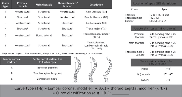
Adolescent idiopathic scoliosis refers to spinal deformity that develops from just before the onset of puberty until the completion of skeletal growth, and the primary goal of treatment is to achieve a well-balanced spine. In the late 1990s, advances in the anatomical understanding of the spine and the development of fixation instruments made posterior pedicle screw insertion feasible, thereby enabling the transmission of powerful corrective forces for deformity correction. Over the subsequent decades, accumulated clinical experience and outcomes have provided a deeper understanding of scoliotic curves and led to the establishment of effective principles for determining the extent of spinal fusion. However, these treatment principles are based on the unique biomechanics and procedural characteristics of scoliosis correction surgery, which can make them difficult to understand without sufficient explanation. In this review, we aim to describe these established treatment principles and surgical processes in detail using schematic illustrations and images. Although these principles will continue to undergo new challenges and validation over time, they will remain a meaningful reference point for those exploring alternative strategies.
Seventy-four-year female patient presented back pain, radiating pain from both posterior thigh and intermittent claudication for two years. Preoperative radiography and MRI demonstrated L2~S1 spinal stenosis. She underwent OLIF and posterior instrumentation L2-S1. At 3 days postoperatively, she presented nausea, abdominal discomfort and showed low SpO2 and drowsy mental state with abrupt vomiting. Abdomen X-ray and CT demonstrated severe paralytic ileus and Chest CT and bronchoscopy demonstrated aspiration pneumonia and ARDS. She transferred to respiratory internal medicine in intensive care unit. She recovered for one month of ICU care and was possible to wheelchair ambulation. Approximately 3.5% of patients undergoing elective spine surgery develop paralytic ileus.
Especially, anterior or lateral access spine surgery, gastroesophageal reflux disease and posterior instrumentation have a high risk of ileus. If patients present nausea, vomiting, abdominal discomfort, constipation, doctor must be evaluated paralytic ileus and treat it by NPO, early ambulation, nasogastric tube and possible pharmacological agents.
Variable posterior surgical techniques for atlantoaxial (C1-2) joint instability (AAI) have been introduced and advanced steadily during the past century. Many surgical techniques using wire or clamp were introduced before 1980s and these surgical approaches provided low biomechanical strength and low fusion rate. After then, screwbased techniques (trans-articular or segmental fixation) were introduced and popularized as an alternative or “gold standard” method. Screw-based methods have recently gained popularity and modified according to their targeted anatomical regions (pedicle, posterior arch, C1 lateral mass, pars inter-articularis and laminar). Each surgical technique has own strength and weaknesses, and their usefulness has been proved through many biomechanical analysis and clinical applications. Advantage and limitation of each surgical technique will be reviewed.
Purpose To analyze the serial changes of the lumbar sagittal alignment from preop. to final follow-up and to evaluate the role of the posterior spinal instrumentation, especially, short level fusion in correction and maintenance of the lumbar sagittal alignment in degenerative lumbar disease.
Materials and Methods Various lumbar sagittal profiles such as lumbar lordosis(LL), lordosis above, within and below instrumentation(LAI, LWI, LBI), horizontal vertebra and sacral inclination were serially measured in 54 patients whose radiographs at preop., intraop., immed. postop. postop. 2wks and final follow up(>1 yr) were completely equipped.
Results Intraop. posture, instrumentation itself and interbody fusion could not increase the LL and LWI sufficiently irrespective of the length of fixation. LWI was decreased compared with preop. values irrespective of length of fixation, while interbody fusion has a great role in maintaining the LWI. Loss of LWI was overcompensated at the segments above instrumentation in 1 or 2 levels fixation while compensation has not occurred in longer fixations.
Conclusions The longer the fixation, the more correction could be obtained. However, maintenance of this correction is more difficult in longer fixations. Prudent consideration should be taken in restoring sufficient lumbar lordosis and maintenance for favorable long term results.

 First
First Prev
Prev

