Study Design This study was a retrospective single-center cohort study.
Purpose This study aimed to compare long-term shunt patency by shunt type (syringo-subarachnoid [SS], syringo-pleural [SP], syringo-peritoneal [SPt]) and disease etiology (post-traumatic, post-infectious, idiopathic) in patients surgically treated for syringomyelia, using Kaplan-Meier survival methodology.
Overview of Literature: Syringomyelia shunting carries a well-documented revision burden, but direct comparative survival data across shunt modalities and etiologic subgroups are scarce, particularly from East Asian centers.
Methods We retrospectively analyzed 42 patients (mean age, 47.6±11.8 years; 57.1% male) who underwent syringomyelia shunting at a tertiary neurosurgical center (January 2000–December 2020) with ≥12 months follow-up. Shunt type was classified as SS (n=12), SP (n=16), or SPt (n=14); etiology as post-traumatic (n=24), post-infectious (n=10), idiopathic (n=6), or hemorrhage/tumor-related (n=2). The primary endpoint was shunt revision surgery. Kaplan-Meier analysis, log-rank testing, and Cox proportional hazards regression were performed.
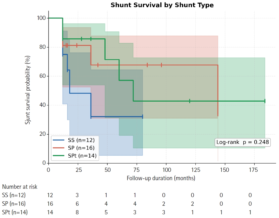
Results Over a median follow-up of 15 months (range, 12 to 184 months), 16 patients (38.1%) underwent shunt revision. Overall 12- and 24-month patency rates were 79.6% and 72.5%, respectively (median shunt survival 72 months). By shunt type, 12-month patency was 75.0% (SS), 81.3% (SP), and 85.7% (SPt); 24-month patency declined to 48.2% for SS while SP and SPt remained at 81.3% and 85.7% (log-rank p=0.248). Post-infectious syringomyelia showed the shortest median shunt survival (36 months) compared with post-traumatic (not reached) and idiopathic (not reached) groups (log-rank p=0.232). No independent predictor of shunt failure was identified on multivariate Cox regression.
Conclusion All three shunting techniques achieve approximately 80% one-year patency. SS may carry a higher long-term occlusion risk. Post-infectious syringomyelia requires closer postoperative surveillance. Larger prospective studies are needed to establish definitive shunt selection criteria.
Objective To investigate the utility of a deep learning model in diagnosing traumatic lumbar fractures on computed tomography (CT) images.
Summary of Background Data: CT scans are widely used as the first choice for detecting spinal fractures in patients with severe trauma. Although CT scans have high diagnostic accuracy, fractures can occasionally be missed.
Recently, deep learning has been applied in various fields of medical imaging.
Methods CT images from 480 patients (3695 vertebrae) who visited a level-one trauma center with lumbar fractures were retrospectively analyzed. The diagnostic results were confirmed by two experienced musculoskeletal radiologists and one experienced spine surgeon using magnetic resonance imaging (MRI). Deep learning networks were employed for diagnosis, with 425 cases used for training and 55 cases for testing. Sensitivity, specificity, accuracy, and the area under the receiver operating characteristic curve (AUROC) were calculated to evaluate diagnostic performance.
Results The model successfully identified 107 out of 129 vertebrae with fractures, achieving a sensitivity of 82.95%, a specificity of 93.24%, an AUROC of 0.936, and an overall accuracy of 88.45%.
Conclusions This study demonstrated that the deep learning model showed high accuracy in diagnosing traumatic lumbar fractures. This approach has the potential to assist spine specialists, radiologists, and trauma care experts.
Further validation is needed to determine its effectiveness in clinical settings.
Introducing a 61-year old woman who was suffering from complicated traumatic thoracolumbar spine fractures, we contemplated the appropriate management algorithm. The Thoracolumbar Injury Classification and Severity score (TLICS) system is the latest and widely used scoring system by spine surgeons for thoracolumbar injuries (TLI). The originator of the system claims for easy application, high reproducibility, and direct link to a clinical decision-making algorithm. However, because of its simple and narrow boundaries, there are many limitations to apply the system in complicated situations. Besides, a fair number of TLI are caused by high velocity traumas, which mostly lead to complicated fractures and other medical conditions. For these reasons, practically, we also consider traditional and former concepts of TLI classifications. Furthermore, new algorithm should be suggested which includes not only the spine morphology and neurological manifestation but also comprehensive medical considerations of the patient.
A 45 year-old male was brought to our hospital with severe back pain and motor, sensory impairment in both lower extremities. He had no underlying diseases including coagulapathy. Motor weakness below both hip joint and decreased sensory below T12 dermatome, voiding dysfunction were examined. The MRI showed a spinal subdural hematoma at the thoracolumbar region, which was extremely rare. Medical treatment was applied without surgical interventions. After two weeks, motor weakness, sensory impairment, and voiding dysfunction were improved. And he returned to his daily activities. We present this case and literature reviews because traumatic spinal subdural hematoma is an extremely rare disease and the condition was treated successfully in conservative manner.


 First
First Prev
Prev