Purpose To evaluate the clinical and radiologic effects of switching from long-term bisphosphonate therapy to romosozumab in an elderly patient with severe osteoporosis and vertebra plana–type severe osteoporotic vertebral collapse, followed by transition to denosumab maintenance therapy.
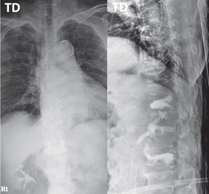
Methods An 85-year-old woman with a history of osteoporotic fractures and prolonged intravenous bisphosphonate therapy presented with an acute L2 compression fracture. Conservative management with a body cast was initiated, and romosozumab was introduced as a switching therapy when anabolic treatment was indicated. Thoracolumbar spine radiographs were obtained at presentation and at 1, 3, 7, and 12 months after injury. Computed tomography was performed at presentation and again at 7 and 12 months to assess fracture consolidation. Bone mineral density (BMD) was measured beginning at 18 months after injury and annually thereafter. Functional assessments were recorded throughout a 30-month follow-up period.
Results During romosozumab therapy, the L2 vertebra plana–type severe osteoporotic vertebral collapse showed marked radiologic improvement, characterized by progressive intravertebral bone fill-in and gradual restoration of trabecular continuity without further loss of height. Serial follow-up CT and MRI demonstrated consolidation of the previously cavitated vertebral body, indicating substantial structural recovery rather than simple stabilization. Clinically, the patient experienced steady improvement in pain and ambulatory capacity. After completing six monthly doses of romosozumab, therapy was transitioned to denosumab. L2 bone mineral density improved from a T-score of –1.7 to –0.9, accompanied by gains in femoral BMD. No additional fragility fractures occurred throughout the follow-up period.
Conclusions Switching from long-term bisphosphonate therapy to romosozumab resulted in improved BMD, progressive vertebral bone fill-in, and stabilization without further collapse in this elderly patient with severe osteoporosis. Although not established as a fracture-healing agent, romosozumab may serve as a practical anabolic option in selected cases, with denosumab maintenance ensuring ongoing skeletal protection.
Background Context: There are few reports of changes in global sagittal alignment and corresponding factors like hand grip strength (HGS) and muscle performance tests to detect changes in global sagittal alignment after surgery for lumbar spinal stenosis (LSS).
Purpose The purpose of the study was to determine whether HGS can be a useful predictive marker of global sagittal alignment changes after decompression with fusion surgery for LSS.
Study Design: This is a retrospective observational study.
Patient Sample: Patients who underwent spine surgery for LSS were included in the present study.
Outcome Measures: Radiological spinopelvic parameters including sagittal vertical axis (SVA), lumbar lordosis (LL), pelvic tilt (PT), pelvic incidence (PI), global tilt (GT), and T1 pelvic angle (T1PA) were assessed. Clinical outcomes parameters like Oswestry Disability Index (ODI), Euro-QOL (EQ-5D), visual analog scale (VAS) scores for back or leg pain were assessed. To assess muscle performance, three functional mobility tests (6-meter walk test, timed up and go test, sit-to-stand test) and HGS were checked.
Materials and Methods A total 91 consecutive patients who underwent spine fusion surgery for LSS were included. 1 year after posterior decompression and fusion surgery, the patients were further classified into four groups according to preoperative and postoperative SVA. We analyzed radiological parameters like SVA, LL, PT, PI, GT, and T1PA. The ODI, the EQ-5D and VAS scores for back or leg pain were recorded as clinical outcomes assessment. To assess muscle performance, SMT, TUGT, STS, and HGS were checked.
Results HGS was significantly correlated with age, postoperative SVA, ODI, EQ-5D and muscle performance test. HGS was related with change of preoperative sagittal alignment 1yr after surgery. Using a receiver operating characteristic (ROC) curve to determine the cutoff values of HGS as predictors of postoperative balanced sagittal alignment according to SVA, cutoff value of HGS demonstrated 19.5 kg with a sensitivity of 82.1% and specificity of 66.7%.
Conclusion Patients with non-balanced sagittal alignment in LSS demonstrated decreased muscle function and muscle strength. If the muscle strength was weak in the group in which the sagittal balance was maintained preoperatively, it could be converted to non-balanced sagittal alignment. Thus, preoperative HGS may be a good predictor of postoperative SVA change.

 First
First Prev
Prev

