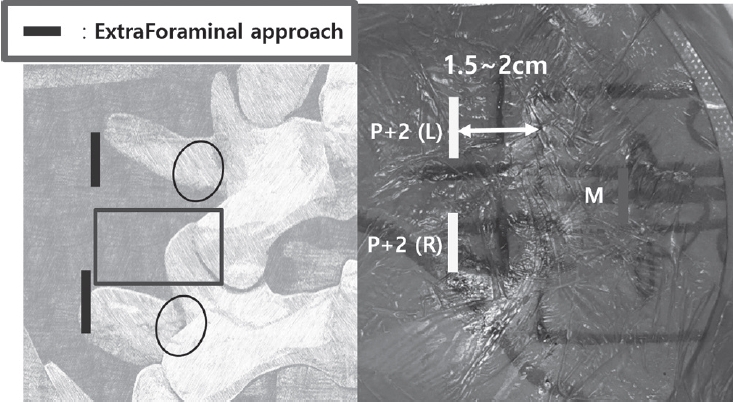
Purpose Revision lumbar surgery following posterior decompression is technically challenging because epidural adhesions and altered anatomy increase the risk of complications during posterior re-entry. Surgical approaches that avoid the previously operated corridor may reduce these risks. Biportal endoscopic lumbar interbody fusion using an extraforaminal approach allows direct neural decompression and interbody fusion through a new surgical corridor, which may be advantageous in revision settings. However, clinical evidence regarding this technique in revision surgery remains limited. To evaluate the clinical and radiological outcomes of biportal endoscopic revision extraforaminal lumbar interbody fusion (BE-REFLIF) performed at lumbar segments previously treated with central decompression.
Materials and Methods This study is Single-center retrospective case series.We retrospectively reviewed 20 consecutive patients who underwent single-level BE-REFLIF as revision surgery after prior central decompression between September 2017 and June 2024. Clinical outcomes were assessed using the visual analogue scale (VAS) for back and leg pain, the Oswestry Disability Index (ODI), and the EuroQol-5D (EQ-5D). Radiological outcomes included disc height, segmental alignment, lumbar lordosis, fusion status, and cage subsidence. Perioperative data and postoperative complications were also analyzed.
Results Significant improvements were observed in all clinical outcome measures during follow-up. Mean VAS scores for back and leg pain and ODI decreased significantly over time (p < 0.001). Radiological analysis demonstrated significant restoration of disc height, improvement in segmental alignment, and maintenance of lumbar lordosis. Solid fusion was achieved in 85% of patients at the final follow-up, and cage subsidence occurred in 25% of cases without the need for reoperation. Perioperative complications included dural tears in 10% of patients, epidural hematoma in 5%, and surgical site infection in 5%, with no instrumentation-related failures.
Conclusions Biportal endoscopic revision extraforaminal lumbar interbody fusion demonstrated favorable clinical and radiological outcomes in patients undergoing revision surgery after previous central decompression. By utilizing an extraforaminal corridor that avoids scarred posterior tissues, BE-REFLIF allows effective direct decompression and interbody fusion with an acceptable complication profile. This technique may represent a viable and less invasive option for selected patients requiring revision lumbar fusion.
Background This study aims to assess the clinical and radiographic outcomes of biportal endoscopic lumbar discectomy compared to microscopic lumbar discectomy in obese patients with lumbar herniated discs. Previous research has established a positive correlation between operation time and estimated blood loss in obese patients undergoing microscopic lumbar discectomy, based on an increase in body mass index. However, no studies have specifically examined the outcomes of biportal endoscopic lumbar discectomy in this patient population. Therefore, this study seeks to fill this research gap and provide valuable insights into the effectiveness of these two surgical approaches for obese patients with lumbar herniated discs.
Methods This retrospective multicenter study analyzed and compared clinical and radiological data from 48 obese patients with a body mass index (BMI) greater than 30 kg/m2 who underwent either microscopic or biportal endoscopic lumbar discectomy. The study assessed clinical outcomes using the visual analog scale (VAS), Oswestry Disability Index (ODI), and EuroQol-5D (EQ-5D) scores. Radiological data were obtained through magnetic resonance imaging (MRI) scans.
Results The study included a total of 48 patients, with 31 patients undergoing microscopic discectomy and 17 patients undergoing biportal endoscopic discectomy. Both groups showed improvements in VAS, ODI, and EQ-5D scores following surgery compared to preoperative scores. However, there was no significant difference in these outcome measures between the two surgical techniques. Although there was a no significant difference in the occurrence of recurrent disc herniation confirmed by postoperative MRI, there was significant difference in the number of patients requiring additional surgery between the two groups.
Conclusions For obese patients with lumbar disc herniation that did not respond to conservative treatment, this study found no significant differences in clinical or radiological outcomes between microscopic and biportal endoscopic surgery methods. However, it is worth noting that the biportal group had a lower incidence of reoperation compared to the microscopic group.
Background When obese patients underwent lumbar discectomy using a microscope, a correlation was found between the operation time and an increase in estimated blood loss according to the increase in body mass index.
However, except for minor complications, there was no difference in postoperative outcomes between obese and normal-weight patients. These are the results of microscopic lumbar discectomy in obese patients, but there are no studies on biportal endoscopic lumbar discectomy. The aim of this study was to compare the clinical and radiographic outcomes of microscopic and endoscopic discectomy in obese patients.
Methods Clinical and radiological data were compared and analyzed in 23 obese patients with a body mass index of >30 kg/m2 who underwent microscopic and biportal endoscopic lumbar discectomy. Clinical data on the visual analog scale (VAS), Oswestry Disability Index (ODI), and EuroQol-5D (EQ-5D) scores were measured, and radiological data were measured using magnetic resonance imaging (MRI).
Results In total, 13 patients who underwent microscopic discectomy and 10 who underwent biportal endoscopic discectomy were enrolled in this study. The VAS, ODI, and EQ-5D scores in both groups improved after surgery compared with those before surgery, although there was no difference between the two groups. Although there was a difference in the incidence of recurrent disc herniation confirmed by MRI after surgery, there was no difference in the number of patients requiring surgery between the two groups.
Conclusions There were no significant clinical or radiological differences in outcomes between microscopic and biportal endoscopic surgery methods.
Objective This study aimed to compare the efficacy of unilateral biportal endoscopic decompression (UBE) and percutaneous endoscopic lumbar discectomy (PELD) in reducing muscle injury by measuring serum levels of creatine phosphokinase (CK) and lactate dehydrogenase (LDH).
Materials and Methods Thirty patients with degenerative lumbar stenosis or a herniated lumbar disc underwent decompression surgery. Among them, 12 patients underwent UBE (experimental group, n=12) and 18 underwent PELD (control group, n=18). CK and LDH were determined at admission and 1, 3, and 5 days after surgery. Pain was measured with a visual analogue scale (VAS).
Results The mean age was significantly higher in the UBE group than the PELD group (63.33±13.50 vs. 49.94±14.79, p<0.035). Mean CK levels were not significantly different at admission. However, at both 3 and 5 days after surgery, CK levels were higher in the UBE group (308.44±153.93 vs. 70.43±40.15, p=0.002; 157.11±91.41 vs. 47.62±23.13, p=0.007). The mean LDH level was higher in the PELD group at 1 day after surgery (152.55±34.69 vs. 199.87±53.78, p=0.027). The operation time was significantly shorter in the PELD group (90.67±39.59 vs. 49.43±14.11, p=0.003).
Conclusions The UBE group had higher CK levels at 3 and 5 days after surgery. The PELD group had a higher LDH level at 1 day after surgery. Therefore, neither procedure is clearly superior in terms of muscle damage.
Spinal subdural hematoma (SDH) is a rare complication after spinal surgery. Only a few cases are reported on spinal SDH following open lumbar spinal decompression or fusion surgery. Moreover, there has been no case report on spinal SDH following percutaneous transforaminal endoscopic lumbar discectomy. We report a case of spinal SDH following endoscopic discectomy, review the literature of this complication and discuss the etiology to it and methods to prevent it. A 63-year-old woman presented with severe radiating pain. Pain was not improved with conservative management. Lumbar magnetic resonance imaging (MRI) was checked and revealed right L3-4 ruptured disc with severe L4 root compression. Percutaneous transforaminal endoscopic decompression was performed and the pain subsided promptly after the endoscopic procedure. On 7th post-operative day, pain on Rt. buttock, anterior thigh was deteriorated severely, more than in pre-operatively. Deteriorated pain was not controlled by oral medications and epidural block. Repeat MRI showed no definite recurrence of disc herniation at decompressed level but spinal SDH, severely compressing cauda equina was seen on T12-sacral area. Spinal SDH is a rare complication following spine surgery, including percutaneous endoscopic surgery. A spine surgeon should be aware of the possibility of spinal subdural hematoma, having severe sequel.
Objective This study aimed to describe the surgical technique in patients with cervical herniated disc treated with unilateral biportal endoscopic spinal surgery.
Materials and Methods Working and viewing portals were created in each unilateral paravertebral area at the target disc level. Under exploring by endoscopic view, effective decompression was possible via safe access to the medial foramen with minimal laminectomy and facetectomy. We evaluated 27 patients, and clinical outcome was analyzed using the visual analogue scale (VAS), Neck disability index (NDI), Macnab criteria, and motor function of involved upper extremity, all assessed before and 3, 6 months post procedure.
Results The VASs for axial neck pain and upper extremity pain decreased from 6.8 to 1.9 and 7.7 to 1.5, respectively, at 6 months post procedure. The NDIs were improved from 45.5 to 13.0 at 6 months post procedure. According to the Macnab criteria, an ‘Excellent’ , ‘Good’ , and ‘Fair’ result was obtained in 55.6%, 29.6%, and 14.8% subjects, respectively.
The motor power of involved upper extremity improved as an approximately one grade on average at 6 months post procedure; 3.9±0.8 to 4.8±0.4.
Conclusions Unilateral biportal endoscopic spinal cervical surgery can be an efficient and safe intervention in patients with cervical herniated disc.
Recently, favorable results of minimally invasive spinal surgery have been reported in comparison to the open decompression or fusion surgery. Biportal endoscopic spine surgery (BESS) has several benefits and Indications for BESS are nearly identical to those for general open spinal surgery. However, it remains a challenging procedure even for an experienced endoscopic surgeon. because it takes a a long operation time while early learning period. If the operation time is prolonged, the advantages of endoscopic surgery are reduced and the incidence of complications can be increased. Therefore, we will investigate the factors affecting the operation time and how to minimize it before and during operation.
Purpose of Study: Purpose of this study is to summarize the technique of UBE surgery in lumbar interbody fusion and review the clinical outcomes and complications of UBE surgery in lumbar interbody fusion.
Materials and Methods Medical databases were searched for the key words of unilateral biportal endoscopic surgery and lumbar spinal stenosis using PubMed from 2005 to the present.
Conclusion UBE spinal surgery is a new technique that can be a feasible alternative and an effective treatment modality for spinal degenerative diseases and can achieve the necessary surgical skills for experienced microscopic surgeons, which is still expanding the indications for lumbar spinal surgery.
Objectives We report the new minimally invasive technique and its clinical results of oblique lumbar interbody fusion (OLIF) combined with central decompression using biportal endoscopic spinal surgery (BESS).
Summary of Literature Review: The OLIF procedure is one of the minimally invasive spine surgeries and is being frequently attempted recently to treat lumbar degenerative disease. It has been reported that it effectively decompresses foraminal stenotic lesions indirectly by inserting a large cage anteriorly, which reduces spondylolisthesis and widens the disc space. However, OLIF has limited effect for severe central canal stenosis, since it cannot achieve direct decompression. Therefore, authors report a new minimally invasive technique of OLIF combined with direct central decompression using BESS for severe central stenosis along with its clinical results as a pilot study.
Materials and Methods For patients who were candidate for fusion surgery due to spondylolisthesis (more than one segment) or foraminal stenosis, authors performed OLIF and central decompression using BESS simultaneously, when the patients had concomitant severe central canal stenosis. From June to December, 2017, 8 patients (16 levels) were enrolled, the operative time, blood loss, complications and clinical results have been evaluated. The clinical results were analyzed by Visual analog scale (VAS) scores, Oswestry disability index (ODI) and Roland Morris Disability Questionnaire (RMDQ) of preoperative, 1month, 3month postoperative and final follow-up.
Results Mean operative time and blood loss were 238.4 minutes and 173.3ml, respectively. In all cases, there were no operative complications, and mean follow-up period was 7.1 months. The mean back VAS, lower extremity VAS, ODI, and RMDQ at the final follow-up were improved from 5.4±2.4 to 2.0±0.9, 7.0±1.1 to 1.6±1.7, 64.2±11.8 to 44.2±10.6, and from 17.5±4.2 to 12.9±4.0.
Conclusion A new combination technique of OLIF and BESS for direct decompression can be regarded as effective alternative procedure to treat the foraminal and central stenotic lesions of lumbar degenerative disease.
The foramen of L5-S1 can develop several degenerative diseases such as extraforaminal lumbar disc herniation, foraminal stenosis with disc height collapse, degenerative or spondylolytic spondylolisthesis, and far-out syndrome.
The floating technique in biportal endoscopic spine surgery (BESS) keeps a certain distance between instruments and spinal structures. 1) This key point makes the floating technique different from conventional endoscopic surgery, which uses the Kambin’s safe triangle as a work zone. The floating view can enable the surgeon to see the structures panoramically, under high magnification: consequently, fine discrimination of their margin and safe manipulation of neural structures can be guaranteed. A certain gap between the floating scope and lesion can permit various instruments, generally used in open spine surgery, to be inserted from the sides with fewer limitations. Extraforaminal or foraminal lesions under the lamina can be reached by avoiding the iliac crest, and total facetectomy, which has the potential of iatrogenic instability, is not required to explore the foraminal structures. However, the floating view can be obstructed by small bleeds from laminectomized bone and/or surrounding vessels. This present article describes the technique and provides tips on how to perform BESS with floating technique safely and successfully for various lesions at the L5-S1 foramen.
Background Owing to its new introduction, there are few documents on pit-falls of biportal endoscopic spine surgery (BESS) clinically. The authors reported etiologies in need of early exploration after BESS for lumbar degenerative diseases and strategies to overcome them.
Methods BESS were performed for lumbar spine diseases (LSDs) by two spine surgeons from December 2013 to March 2016. Postoperative MRI was checked for all cases and following-up MRIs in the case in need of revision surgery within six months after the first surgery due to pain intolerable, sustained or recurred. The complicated cases were reviewed and classified as radiographic and operative findings to reveal the main reasons for early explorations.
Results The 562 cases (M:295, F:267, Age 58.5±14.1 yrs, 20~88 yrs) included lumbar disc herniation (LDH) (255 cases), extraforaminal disc herniation (22 cases), spinal stenosis (218 cases), degenerative spondylolisthesis (27 cases), revision surgery after recurred disc herniation or restenosis after open surgery (24 cases), juxtafacet cyst (11 cases), adjacent segment stenosis with fusion surgery (3 cases), and spondylolytic spondylolisthesis (2 cases). Early explorations were needed in 43 cases (7.7%) at 26.1±31.5 days after the initial operations. Causative etiologies were listed as recurred LDH (12 cases, 27.9%), remnant stenosis (7 cases, 16.3%), remained ruptured disc fragment (6 cases, 14.0%), root edema (5 cases, 11.6%), synovitis (4 cases, 9.3%), hematoma (3 cases, 7.0%), dura tear (2 cases, 4.7%), recurred stenosis (2 cases. 4.7%), wrong level (1 case, 2.3%) and postoperative fungal infection (1 case, 2.3%).
Thirty-one cases (72.1%) were revised within 4 weeks and most conditions (40 cases, 93.0%) were improved after early exploration using BESS. Two cases of dura tear were conversed to open repair. One case of fungal infection was suspected to related with the patient’s medical illness including long-term steroid use for chronic lung disease with pulmonary fibrosis and Diabetes mellitus.
Conclusions Preoperative planning should be prepared carefully to decrease early exploration. It was helpful to comparing MRIs immediately postoperative and early following-up to find the reasons. Don’t hesitate to explore the operated site again using BESS, because most etiologies are supposed to be controlled by early exploration without need of converting to open surgery except in the case of dura tear in need of dural repair.
Since its introduction in 1980s, Laser therapy, a minimally invasive, outpatient, effective, and relatively safe procedure for spine disease, has established itself world-wide. percutaneous laser disc decompression (PLDD) was mainly applied to treat lumbar disc herniation with a satisfactory efficacy. Recently, laser therapy has been used for many cases of lumbar stenosis and cervical disease.In this article, advantages and disadvantages of laser will be discussed, and technical and clinical aspects of current applications will presented. Such applications include Percutaneous lumbar disk decompression (PLDD), Percutaneous endoscopic laser annuloplasty(PELA), Epiduroscopic laser neural decompression(ELND), Laser disk decompression in cervical disk diseases.

 First
First Prev
Prev

