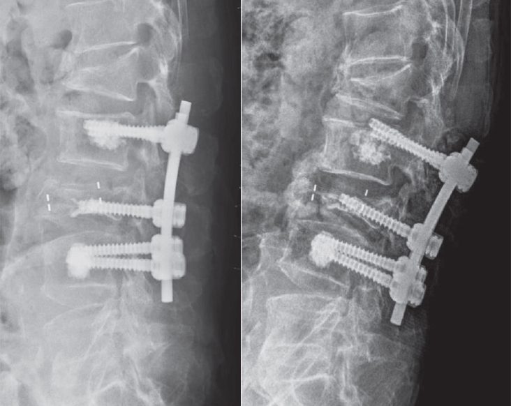
Objective This study aimed to evaluate the stability of cement-augmented pedicle screws in patients with osteoporosis of the thoracolumbar spine, with a focus on reducing mechanical failures compared with non-augmented screws.
Methods A retrospective analysis was conducted on 119 patients who underwent thoracolumbar fusion surgery between 2011 and 2022. The incidence of mechanical failures—including pull-out, screw loosening, and cage protrusion—was compared between patients treated with cement-augmented pedicle screws and those without augmentation.
Results Cement augmentation was associated with a significant reduction in overall mechanical failures. The incidence of mechanical failure was significantly lower in the cement-augmented group compared with the non-augmented group (20.4% vs. 41.4%, p=0.018). Although individual complications such as pull-out, cage protrusion, and screw loosening were less frequent in the cement-augmented group, these differences were not statistically significant. However, the overall reduction in mechanical failures was statistically significant. Fusion rates were higher in the cement-augmented group than in the non-augmented group, although the difference was not significant (79.6% vs. 70.0%, p=0.337). Importantly, patients without mechanical failures had significantly higher fusion rates than those with failures (82.5% vs. 56.41%, p=0.0048).
Conclusions Cement-augmented pedicle screws significantly reduce the risk of mechanical failures in thoracolumbar fusion surgery for patients with osteoporosis. Mechanical stability strongly influences fusion success, highlighting the importance of preventing mechanical failure to optimize surgical outcomes. These findings support cement augmentation as an effective strategy to enhance the durability of pedicle screw fixation and should be considered in surgical planning for patients with osteoporosis.
Purpose To evaluate the clinical and radiologic effects of switching from long-term bisphosphonate therapy to romosozumab in an elderly patient with severe osteoporosis and vertebra plana–type severe osteoporotic vertebral collapse, followed by transition to denosumab maintenance therapy.
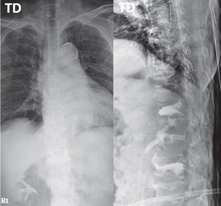
Methods An 85-year-old woman with a history of osteoporotic fractures and prolonged intravenous bisphosphonate therapy presented with an acute L2 compression fracture. Conservative management with a body cast was initiated, and romosozumab was introduced as a switching therapy when anabolic treatment was indicated. Thoracolumbar spine radiographs were obtained at presentation and at 1, 3, 7, and 12 months after injury. Computed tomography was performed at presentation and again at 7 and 12 months to assess fracture consolidation. Bone mineral density (BMD) was measured beginning at 18 months after injury and annually thereafter. Functional assessments were recorded throughout a 30-month follow-up period.
Results During romosozumab therapy, the L2 vertebra plana–type severe osteoporotic vertebral collapse showed marked radiologic improvement, characterized by progressive intravertebral bone fill-in and gradual restoration of trabecular continuity without further loss of height. Serial follow-up CT and MRI demonstrated consolidation of the previously cavitated vertebral body, indicating substantial structural recovery rather than simple stabilization. Clinically, the patient experienced steady improvement in pain and ambulatory capacity. After completing six monthly doses of romosozumab, therapy was transitioned to denosumab. L2 bone mineral density improved from a T-score of –1.7 to –0.9, accompanied by gains in femoral BMD. No additional fragility fractures occurred throughout the follow-up period.
Conclusions Switching from long-term bisphosphonate therapy to romosozumab resulted in improved BMD, progressive vertebral bone fill-in, and stabilization without further collapse in this elderly patient with severe osteoporosis. Although not established as a fracture-healing agent, romosozumab may serve as a practical anabolic option in selected cases, with denosumab maintenance ensuring ongoing skeletal protection.
Purpose To compare the 3-month outcomes of romosozumab and percutaneous vertebroplasty in patients with acute osteoporotic vertebral compression fractures (OVCFs).
Background Vertebroplasty provides rapid pain relief in acute OVCFs but carries risks such as cement leakage and adjacent fractures. Romosozumab, an anti-sclerostin monoclonal antibody, promotes bone formation and reduces fracture risk; however, its effectiveness in acute OVCFs remains unclear.
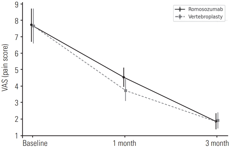
Material and Methods: This retrospective study included 84 patients with MRI-confirmed acute OVCFs treated between January 2022 and December 2024. Patients received either monthly subcutaneous romosozumab injections (n=52) or vertebroplasty followed by weekly oral alendronate (n=32). All received daily calcium (500 mg) and vitamin D₃ (1,000 IU). Clinical outcomes were assessed using the Visual Analogue Scale (VAS) and Oswestry Disability Index (ODI), and radiographic changes were evaluated based on anterior vertebral body height at baseline, 1 month, and 3 months.
Results Both groups showed significant improvements in VAS and ODI scores at 1 and 3 months, with no significant differences between them. Vertebral height changes were also comparable.
Conclusions Romosozumab-based conservative therapy may be a viable non-invasive alternative to vertebroplasty in treating acute OVCFs, offering similar short-term clinical and radiographic outcomes.
Purpose The impact of skeletal muscle mass and bone mineral density (BMD) on frailty after osteoporotic vertebral fractures (OVFs) remains unclear. This study aimed to assess the interplay between frailty, skeletal muscle mass, and bone mineral density in OVFs.
Materials and Methods A total of 66 patients with osteoporotic vertebral compression fractures were enrolled. We collected clinical and radiological data, including age, body mass index (BMI), frailty index, and parameters such as lumbar lordosis, thoracic kyphosis, skeletal muscle mass, and BMD. We then analyzed the relationships between frailty and these variables.
Results The mean age, BMI, BMD T-score, skeletal muscle mass, and frailty index were 78.0±7.8 years, 22.3±3.3 kg/ m², -3.59±0.96, 37.84±6.24 kg, and 2.59±1.08, respectively. Of the 66 patients, 14 (21.1%) were frail prior to fracture, while 37 (56.1%) were frail after fracture, indicating a worsening frailty status. Specifically, 23 patients (34.8%) transitioned from pre-frail to frail following their fracture and had both lower BMD (T-score: -3.7±0.93) and lower skeletal muscle mass (35.74±3.83 kg). Frailty was negatively correlated with BMD (r=-0.28, p=0.02), while BMD was positively correlated with skeletal muscle mass (r=0.29, p=0.02). OVFs were positively correlated with frailty (r=0.33, p=0.01), especially in terms of fatigue (r=0.31, p=0.01) and ambulation (r=0.21, p=0.01).
Conclusions In patients with osteoporotic vertebral fractures, decreased muscle mass and low BMD appear to exacerbate frailty. Furthermore, frailty may be both a contributing and a resulting factor in the development of osteoporotic vertebral fractures.
Introduction We describe the complications that can occur after percutaneous vertebroplasty using bone cement for osteoporosis vertebral compression fracture.
Main subject: The most common complication of percutaneous vertebroplasty is the leakage of bone cement.
Leakage of bone cement has been reported variously and could leak into the spinal or neural foramen, adjacent intervertebral disc and soft tissues around the spine, and venous systems. The most serious complications are neurologic symptoms due to spinal cord and nerve root compression and complications associated with death due to heart and pulmonary embolism. In addition, recompression fracture or adjacent vertebral compression fracture might occur and various treatment methods have been proposed.
Conclusion The complications that can occur after percutaneous vertebroplasty have been reported variously, including neurologic deficits due to the leakage of bone cement and lung and heart embolism. In addition, there is a possibility of recompression fracture or adjacent compression fracture. Therefore, you should be careful about percutaneous vertebroplasty. Finally, patients with many risk factors regarding complications of vertebroplasty would need close observation and follow-up.
When conservative treatment fails in the treatment of osteoporotic vertebral compression fractures, a minimally invasive procedure, such as percutaneous balloon kyphoplasty or vertebroplasty is performed. Among these, balloon kyphoplasty is known as an advantageous method for lower risk of cement leakage and greater correction effect of kyphosis and better sagittal balance correction. However, there are reports of various complications during and after procedure, and sometimes result in serious consequences. This paper reviews with previous literatures about the complications related to balloon kyphoplasty.
Percutaneous vertebroplasty and balloon kyphoplasty are both safe and effective procedures in case of patients with osteoporotic vertebral compression fractures. The authors have already reported a new technique called lordoplasty using polymethylmethacrylate to manage vertebral osteoporotic compression fractures. The purpose and indication of lordoplasty do not differ from that of percutaneous vertebroplasty or balloon kyphoplasty. However, there are advantages of lordoplasty in terms of restoration of the wedge and kyphotic angle and cost-effectiveness compared with the other procedures mentioned above. For the advantages of lordoplasty, authors thereby introduce the detailed procedure of lordoplasty.
Purpose Osteoporosis is an age-related systemic skeletal disease characterized by low bone mass and microarchitectural deterioration of bone contents, with a consequent increase in bone fragility. In severe osteoporosis progressive collapse of multiple vertebrae is and unsolved problem. Medical treatment appears to be too slow to prevent the course. Recently, there are some reports on the results of the percutaneous vertebroplasty (VP) in treating the multi-level osteoporotic vertebral compression fractures (VCFs). we reviewed painful multi-level osteoporotic VCFs treated by percutaneous VP and assess the efficacy and safety of multiple percutaneous cement VP in the treatment of multi-level osteoporotic VCFs.
Materials and Methods From January 2008 to August 2010, the clinical cases and radiographic records were reviewed retrospectively for 28 patients treated for the multi-level painful osteoporotic VCFs by percutaneous cement VP.
Initially radiography and MRI of the spine were performed. Spine radiographs were repeated at post-operation, 1,3 months and final follow-up. The patient’s outcomes of demographic, clinical, radiologic and procedural data were analyzed and assessed using self-report and physiological measures. A t-test was used for means of VAS, anterior vertebral height and kyphotic angle. Statistical analysis was performed with the SPSS(Version 15.0.1, Chicago, Illinois). The p-values of < 0.001 were deemed significant.
Results The back pain recorded using the VAS improved significantly in all cases, from 7.7±1.0(6-10), points preoperatively to 2.0±0.7(1-3) points postoperatively (p<0.001) and then 2.8±0.8(1-4) points at the follow-up (p<0.001).
The anterior heights increased from 17.40±4.98 to 21.02±5.36 after VP procedures (p<0.001) and finally 19.49±5.28 (p<0.001). The kyphotic angle was 12.58º preoperatively and improved to 4.39º postoperatively, but kyphotic deformities became worse in 12.80º.
Conclusion The vertebroplasty for patients with multiple osteoporotic vertebral compression fractures may improve pain and can be effective for preventing adjacent fractures, restoration of vertebral height and maintenance of sagittal alignment. Patients with multiple osteoporotic compression fractures have many comorbidity, the surgeon should be conscious to all procedure.

 First
First Prev
Prev

