Impact of Percutaneous Vertebroplasty on Vertebral Height and Sagittal Alignment in Thoracolumbar Osteoporotic Compression Fractures: A Retrospective Observational Study
1)Department of Neurosurgery, International St. Mary’s Hospital, Catholic Kwandong University College of Medicine, Incheon, Korea
2)Department of Neurosurgery, Daegu Catholic University School of Medicine, Daegu, Korea
Corresponding author: Kwang-Ryeol Kim, M.D. Department of Neurosurgery, Daegu Catholic University School of Medicine, 33 Duryugongwon-ro 17-gil, Nam-gu, Daegu 42472, Korea TEL: +82-53-650-4539, FAX: +82-53-650-4932, E-mail: ianremedios@cu.ac.kr
• Received: December 19, 2025 • Revised: February 4, 2026 • Accepted: February 10, 2026
This is an open access article distributed under the terms of the Creative Commons Attribution Non-Commercial License (https://creativecommons.org/licenses/by-nc/4.0/) which permits unrestricted non-commercial use, distribution, and reproduction in any medium, provided the original work is properly cited.
This study aimed to evaluate whether percutaneous vertebroplasty (PVP) contributes to vertebral height restoration and sagittal alignment correction in osteoporotic vertebral compression fractures (OVCF), focusing on thoracolumbar junction fractures.
Methods
A retrospective review of 40 patients with single-level OVCF at T10–L2 treated with PVP was performed. Vertebral heights (anterior, middle, and posterior) and sagittal alignment (thoracic kyphosis, lumbar lordosis, sagittal vertical axis, and segmental Cobb's angle) were measured preoperatively, at 3 months, and at 6 months. Clinical outcomes included visual analog scale and EuroQol-5 Dimensions.
Results
Significant pain relief and improvement in quality of life were observed at 6 months postoperatively. Vertebral height restoration, particularly in the anterior and middle portions, was noted at 3 months; however, partial loss of the restored height occurred by 6 months. Most sagittal alignment parameters showed no significant postoperative change, although lumbar lordosis significantly increased, resulting in a reduced pelvic incidence–lumbar lordosis mismatch.
Conclusion
PVP provides meaningful clinical improvement in thoracolumbar OVCFs and offers early vertebral height restoration; however, this radiologic benefit is not sustained over time. While limited improvement in lumbar lordosis was observed, PVP does not substantially correct global sagittal alignment. These findings suggest that PVP should be considered primarily a pain-relieving and stabilizing procedure rather than a deformity-correcting intervention.
Osteoporotic vertebral compression fracture (OVCF) is among the most common fragility fractures in the aging population, with more than half of individuals over 80 years of age experiencing at least one fracture during their lifetime. These fractures often lead not only to acute mechanical back pain but also to progressive vertebral body collapse, segmental kyphosis, and global sagittal imbalance, ultimately reducing mobility, independence, and quality of life.1,2) The thoracolumbar junction (T10–L2) is particularly vulnerable to osteoporotic fractures because it constitutes a biomechanical transition zone between the rigid, kyphotic thoracic spine and the more flexible, lordotic lumbar spine.3) Accordingly, OVCFs most frequently occur in this region, and fractures at T10–L2 tend to cause more pronounced deformity and instability than those at other spinal levels.4,5)
Conservative management remains the first-line treatment for most OVCFs. Standard care involves 1–2 weeks of absolute bed rest with back support, followed by using a rigid thoracolumbar orthosis.6,7) However, pain control may be insufficient in a considerable proportion of patients, especially those with severe collapse or progressive kyphosis. In such cases, surgical options including percutaneous vertebroplasty (PVP) or kyphoplasty (KP) are considered.8,9)
Previous studies have shown heterogeneous results, partly due to variation in fracture levels, inclusion of multilevel fractures, differences between PVP and KP, inconsistent follow-up periods, and the confounding effect of osteoporosis pharmacotherapy.10-12) There have even been reports that PVP has no significant effect in pain control.13,14) On the other hand, there are also reports that it is helpful not only in controlling early pain but also in controlling chronic pain and reducing mortality.15-18)
Although PVP is widely accepted as an effective method for rapid pain relief, its impact on vertebral height restoration and correction of segmental kyphosis remains controversial.19,20) Thus, the true radiologic and clinical impact of PVP in OVCFs, particularly at the anatomically critical thoracolumbar junction remains unclear.
Therefore, this study evaluates whether PVP meaningfully restores vertebral height and improves global or segmental alignment in thoracolumbar OVCF.
Methods
1. Study population
This retrospective observational study included patients who underwent PVP or KP for OVCFs at a single institution between March 2020 and February 2023. A total of 157 consecutive patients were initially identified. To minimize confounding effects on radiologic outcomes, patients were excluded if they had multilevel fractures or previous adjacent-level fractures (n=27), underwent KP (n=25), had a follow-up duration of less than 6 months or lacked follow-up whole-spine standing radiographs (WSXR) (n=17), received anabolic osteoporosis agents such as romosozumab or teriparatide (n=15), or had pathologic fractures due to metastatic disease or Kümmell’s disease (n=14).
After applying these exclusion criteria, 59 patients who underwent PVP for a single-level OVCF were eligible for analysis. To focus specifically on fractures occurring at the biomechanically vulnerable thoracolumbar junction, only patients with fractures at T10–L2 were included in the final study population (n=40). Patients with mid-thoracic (T6–8, n=9) or lower lumbar (L3–5, n=10) fractures were excluded from the primary analysis.
2. Procedural technique
All PVP procedures were performed by a single experienced spine surgeon under biplanar fluoroscopic guidance.
All procedures were performed with the patient in the prone position under sterile conditions. After routine skin preparation and surgical draping, the target pedicle was identified using a C-arm fluoroscope in the anteroposterior (AP) view. The superolateral margin of the target pedicle was infiltrated with 1% lidocaine to achieve local anesthesia of the skin and paraspinal muscles. A 0.5-cm skin incision was made using a No. 15 blade, and a Jamshidi needle was advanced through the pedicle under fluoroscopic guidance.
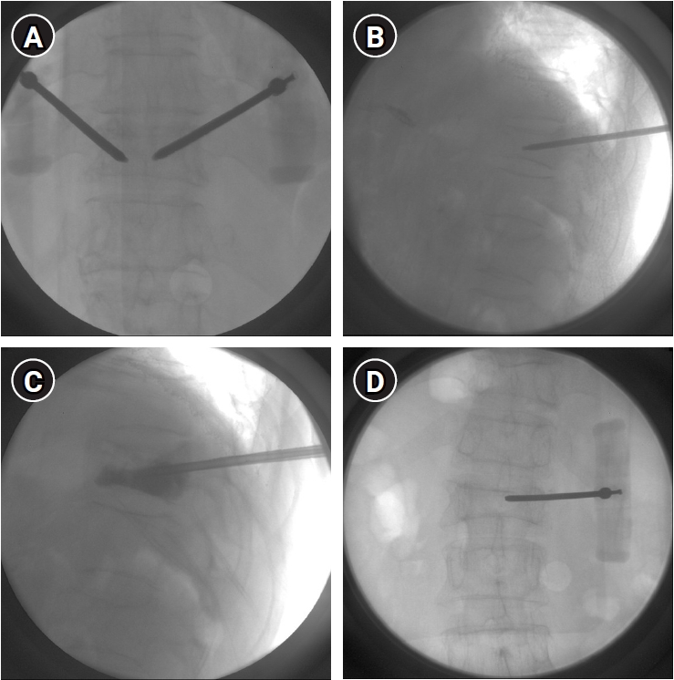
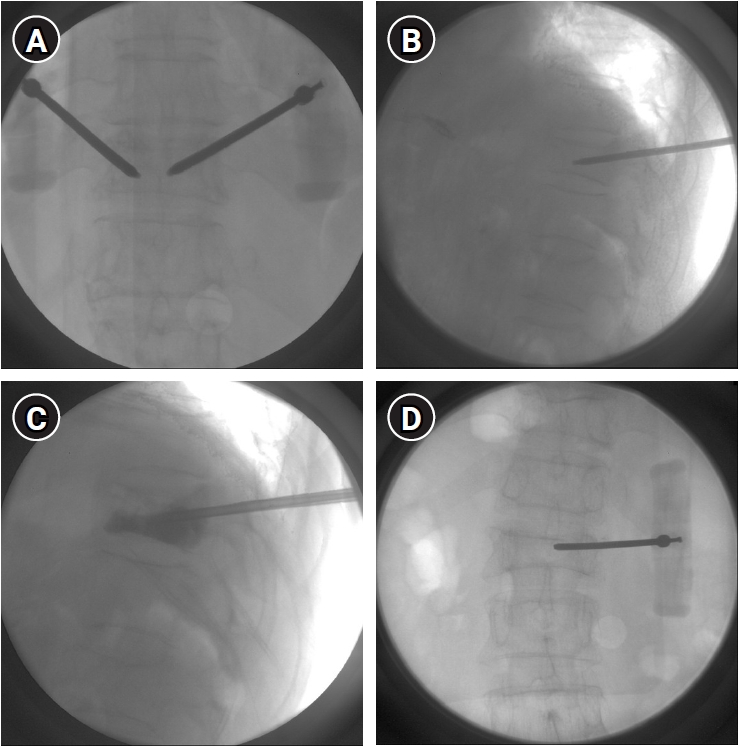
Needle advancement was monitored using alternating AP and lateral fluoroscopic views. Once the needle tip was confirmed to be positioned within the anterior one-third of the vertebral body on the lateral view, polymethylmethacrylate (PMMA) cement was prepared and injected during the doughy phase under continuous fluoroscopic monitoring. Cement injection was terminated immediately upon adequate filling of the vertebral body or when any sign of cement leakage was detected (Fig. 1).
For the unilateral transpedicular approach, the needle tip was directed slightly more medially compared with the bilateral approach to facilitate cement distribution across the midline.21) In addition, a relatively lower-viscosity PMMA cement was used to promote bilateral intravertebral spread. If adequate cement distribution to the contralateral side was not achieved during injection via the unilateral approach, an additional transpedicular procedure on the contralateral side was considered to ensure sufficient vertebral body filling and stabilization.
A unilateral transpedicular approach was used in 22 cases, while a bilateral transpedicular approach was employed in 18 cases, depending on fracture morphology and cement distribution.
Postoperatively, patients were maintained on absolute bed rest for 4 hours and were subsequently mobilized with a rigid thoracolumbar orthosis on the same or following day. The brace was worn during ambulation and daily activities for 3 months after the procedure.
3. Clinical assessment
Clinical outcomes were evaluated at baseline (preoperatively) and at 6 months postoperatively. Back pain intensity was assessed using the visual analog scale (VAS), and health-related quality of life was evaluated using the EuroQol-5 Dimensions (EQ-5D) index. Changes in clinical outcomes were analyzed in relation to radiologic findings to assess the association between symptomatic improvement and structural changes following PVP.
4. Radiologic assessment
Radiologic evaluations were performed at three times: preoperatively (within 1 week before PVP), and at 3 months and 6 months postoperatively. WSXR were obtained at each time point and used for all measurements.
Vertebral body height was assessed at the fractured level by measuring anterior, middle, and posterior vertebral heights. Measurements were recorded as absolute values and, when applicable, expressed as a percentage of the estimated normal height based on adjacent intact vertebrae.
Sagittal alignment parameters included thoracic kyphosis (TK) measured as the Cobb angle from T5 to T12, lumbar lordosis (LL) measured from L1 to S1, sagittal vertical axis (SVA), and the segmental Cobb angle (SCA) at the fractured level representing local kyphosis. All radiologic parameters were independently measured by two spine surgeons using a Picture Archiving and Communication System–based digital measurement system, and the mean of the two measurements was used for statistical analysis.
5. Statistical analysis
Continuous variables are presented as mean±standard deviation, and categorical variables are expressed as frequencies and percentages. Normality of continuous variables was assessed using the Shapiro-Wilk test. Changes in clinical outcomes (VAS and EQ-5D scores) and radiologic parameters (sagittal alignment variables) before and after PVP were analyzed using paired t-tests. For variables that did not meet the assumption of normality, the Wilcoxon signed-rank test was applied. Changes in vertebral body height (anterior, middle, and posterior) across three points (preoperative, 3 months, and 6 months) were analyzed using one-way repeated-measures analysis of variance (ANOVA). When a significant overall effect was detected, post hoc pairwise comparisons were performed with Bonferroni correction. Statistical analyses were performed using SPSS software version 19.0 (IBM Corp., Armonk, NY, USA). All tests were two-tailed and a p-value < 0.05 was considered statistically significant.
6. Ethics statement
This retrospective study was approved by the Institutional Review Board of our institution (IRB No. IS25RISI0074) and was conducted in accordance with the principles of the Declaration of Helsinki. Because of the retrospective design of the study and the use of de-identified data, the requirement for informed consent was waived by the Institutional Review Board.
Results
1. Patient characteristics
The patient cohort in this study comprised nine men and 31 women, with median age of 82.20 years (range, 39 to 107 years). The L1 vertebral level was the most commonly affected level, with 16 patients (40.0%), followed by L2 in 10 patients (25.0%) and T12 in eight patients (20.0%). Mean injected PMMA volume was 5.53 mL (range, 3 to 9 mL), mean time to surgery 9.58 days (range, 1 to 47 days), and mean operative time 29.73 minutes (range, 15 to 50 minutes). Bone mineral density T-scores were −3.19 (lumbar spine) and −3.02 (femoral neck) (Table 1).
2. Clinical outcomes
The mean VAS scores for back pain before PVP were 7.80±1.47. At 6 months after PVP, the mean VAS scores decreased to 3.30±1.47. The VAS score decreased by approximately 4.5 points before and after the procedure.
The mean EQ-5D score was 0.51±0.23 before PVP. It increased by 0.21 to 0.72±0.18 after PVP (Table 2).
3. Radiologic outcomes
1) Vertebral height
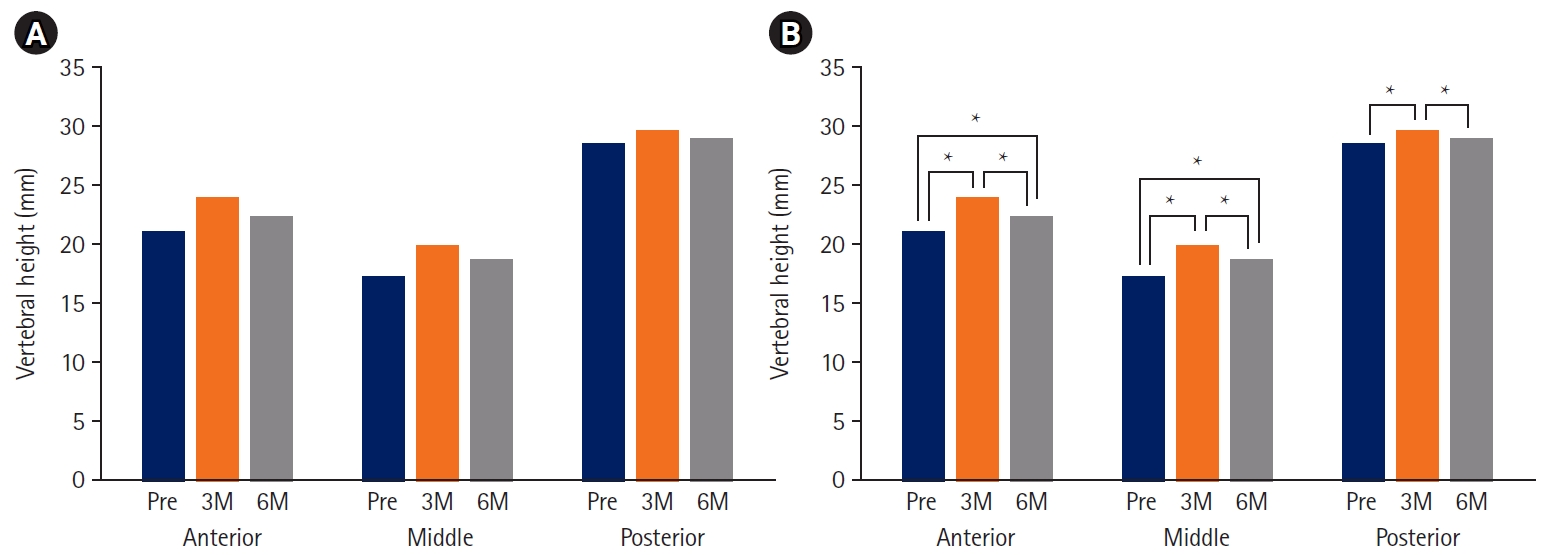
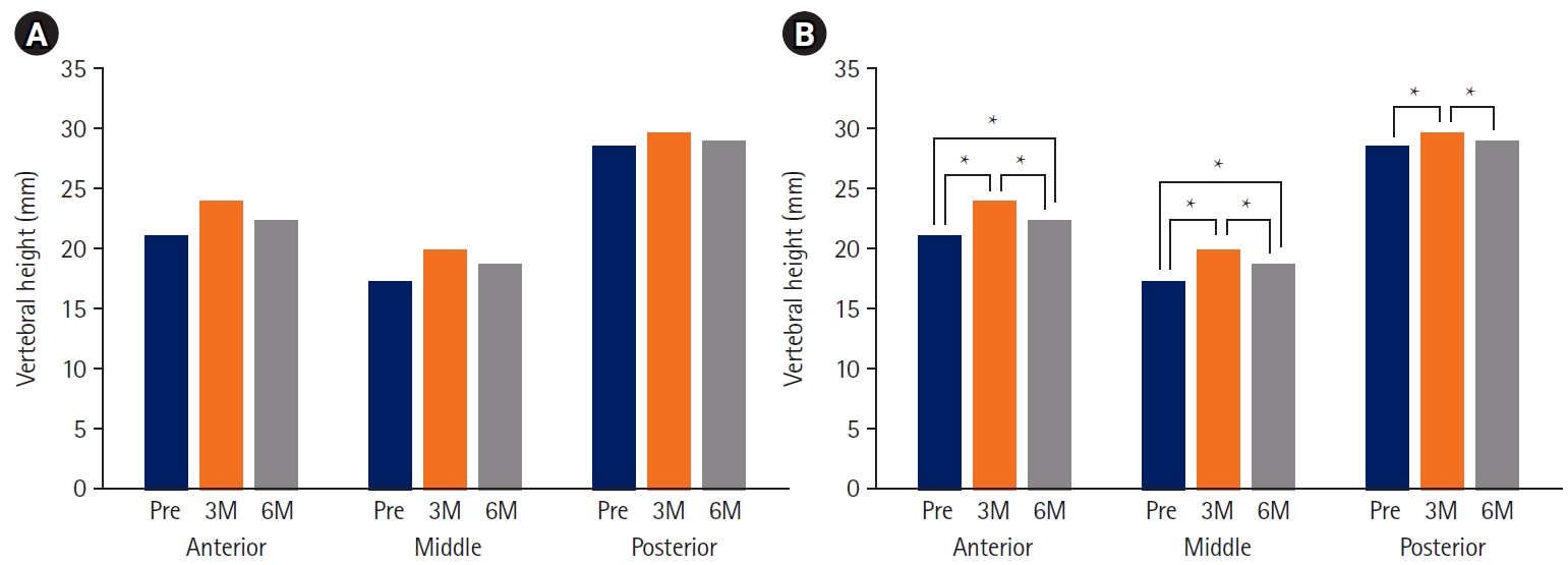
Radiographic analysis demonstrated early improvement in vertebral body height following PVP. At 3 months postoperatively, the mean anterior vertebral height increased from 21.13 mm preoperatively to 23.94 mm, and the mean middle height increased from 17.23 mm to 19.90 mm. A modest increase was also observed in the posterior vertebral height, from 28.55 mm to 29.68 mm.
However, at the 6-month follow-up, partial loss of the restored vertebral height was observed. The mean anterior height decreased to 22.35 mm, and the middle height decreased to 18.64 mm, while the posterior height slightly decreased to 28.94 mm. Although vertebral heights at 6 months remained higher than preoperative values, the initial postoperative gains were not fully maintained over time (Fig. 2).
2) Sagittal alignment
Assessment of sagittal alignment revealed minimal changes following PVP. The mean TK angle showed a slight decrease from 29.84° preoperatively to 29.53° at follow-up. LL demonstrated a small increase from 29.84° to 31.13°. The SVA decreased marginally from 66.45 mm to 65.11 mm. The SCA at the fractured level increased slightly from 7.11° to 7.78°. Among these, LL significantly increased and pelvic incidence minus lumbar lordosis (PI–LL) significantly decreased (Table 3).
4. Correlation between radiologic and clinical outcomes
Correlation analysis was performed to determine whether the degree of vertebral height restoration influenced clinical improvement. No significant correlation was observed between the restoration of anterior, middle, or posterior heights and changes in VAS or EQ-5D scores at any follow-up interval (Table 4).
5. Subgroup analysis based on baseline sagittal balance
To further investigate whether the preoperative sagittal profile influences radiologic outcomes, patients were categorized into subgroups based on SVA and PI–LL mismatch. For SVA, a cutoff of 50 mm was used as it represents the threshold for normal sagittal balance.22) Regarding PI–LL mismatch, while a 10° threshold is often considered the upper limit of normal, applying this cutoff resulted in a significant disproportion in sample sizes between groups. Therefore, a cutoff of 20° was adopted to represent severe sagittal imbalance, ensuring a more balanced distribution for statistical comparison.23) Based on these criteria, subgroup analysis was performed to compare changes in TK and SCA (Table 5).
Discussion
The present study demonstrates that PVP provides significant clinical improvement in patients with thoracolumbar OVCFs. Meaningful reductions in pain scores and improvements in health-related quality of life were observed at 6 months postoperatively. Radiologic evaluation revealed modest and transient restoration of vertebral body height, which was not fully maintained over time. With respect to sagittal alignment, most global and segmental parameters, including TK, SVA, and SCA, showed no substantial postoperative change; however, LL demonstrated a significant postoperative increase, accompanied by a corresponding reduction in the PI–LL mismatch. Despite these limited alignment-related changes, the overall findings suggest that the primary benefits of PVP are derived from pain relief and mechanical stabilization rather than from durable anatomic restoration or global sagittal realignment.
The substantial pain relief observed in this study is consistent with previous reports supporting the analgesic efficacy of PVP. The mechanism underlying this improvement is likely related to mechanical stabilization of the fractured vertebral body, reduction of micro-motion at the fracture site, and thermal or chemical effects of PMMA cement.24,25) Our correlation analysis further reinforces this, as no significant association was found between the magnitude of height restoration and clinical outcomes (Table 4). Importantly, the observed clinical benefits occurred despite the absence of meaningful sagittal realignment, reinforcing the concept that pain relief after PVP is not dependent on restoration of vertebral geometry or global spinal balance.26,27)
With respect to vertebral height, our results demonstrate a clear early postoperative increase, particularly in the anterior and middle portions of the vertebral body. However, this restoration was only partially maintained for 6 months, indicating gradual settling of the fractured vertebra over time. These findings align with biomechanical and clinical studies suggesting that while PVP increases local stiffness, it does not reconstitute the trabecular architecture or load-bearing capacity of osteoporotic cancellous bone. Consequently, progressive collapse may occur even after technically successful cement augmentation.28,29) This observation is particularly noteworthy when considering the natural course of untreated OVCFs. While untreated fractures typically undergo progressive and irreversible collapse, our data showed an initial significant height restoration followed by only partial loss. This suggests that while PVP may not permanently 'fix' the anatomy to a pre-fracture state, it potentially alters the natural history of vertebral collapse by providing early mechanical stabilization.
Despite early height restoration, sagittal alignment parameters, including TK, LL, SVA, and SCA, remained largely unchanged. Several factors may account for this observation. First, the thoracolumbar junction is subject to high transitional biomechanical stress, limiting the impact of localized vertebral height changes on global alignment.30) Second, PVP does not restore disc height, ligamentous tension, or posterior column integrity, all of which play critical roles in sagittal balance.31) Finally, the magnitude of vertebral height restoration observed in this study was relatively small and likely insufficient to translate into measurable changes in global or segmental alignment. Furthermore, our subgroup analysis demonstrated that even in patients with significant baseline sagittal imbalance (SVA ≥50 mm or PI–LL ≥20°), the corrective impact of single-level PVP on local or global alignment remained negligible (Table 5).
From a clinical perspective, these findings indicate that PVP should be regarded primarily as a procedure for pain control and fracture stabilization rather than a deformity-correcting intervention. Although early vertebral height restoration may be observed, it is not reliably sustained, and meaningful correction of kyphotic deformity or sagittal imbalance should not be expected. Accordingly, patient counseling should emphasize the symptomatic benefits of PVP while setting realistic expectations regarding radiologic and alignment outcomes, particularly in fractures involving the thoracolumbar junction.
Despite the clinical relevance of our findings, this study has several limitations that should be acknowledged. First, the follow-up duration was relatively short, which may be insufficient to fully capture long-term changes in vertebral height, sagittal alignment, and fracture progression after PVP. Second, the most significant limitation is the absence of a control group treated conservatively. Without a comparative cohort, it is difficult to definitively distinguish the therapeutic effects of PVP from the natural radiographic progression of OVCFs. However, the rigorous inclusion of only single-level thoracolumbar junction fractures allowed for a more focused observation of the procedure’s specific impact on this biomechanically sensitive region. Future prospective randomized controlled trials comparing PVP with conservative management are necessary to further elucidate the isolated impact of cement augmentation on spinal kinematics. Third, radiologic analysis was based primarily on lateral WSXRs, and AP radiographic analysis was not performed. As a result, the lateral distribution of PMMA cement within the vertebral body could not be assessed, and potential associations between cement dispersion patterns and radiologic or clinical outcomes could not be analyzed. These limitations suggest that the findings should be interpreted with caution and underscore the need for future studies with longer follow-up periods, comparative study designs, and more comprehensive radiographic evaluation.
In conclusion, while PVP for single-level thoracolumbar OVCFs provides significant pain relief and immediate restoration of vertebral height, its impact on global sagittal alignment appears limited. Our findings suggest that PVP may contribute to local segmental stabilization rather than substantial correction of global sagittal balance. Further large-scale, prospective controlled trials are warranted to confirm these observations.
NOTES
Author contributions
Conceptualization: KRK. Data curation: JO, KRK. Formal analysis: GJP, KRK. Investigation: JO, KRK. Funding acquisition: KRK. Methodology: GJP, KRK. Project administration: KRK. Resources: JO, KRK. Software: JO, GJP. Supervision: KRK. Validation: JO, KRK. Visualization: GJP, KRK. Writing – original draft: JO, KRK. Writing – review & editing: KRK.
Conflict of interest
Kwang-Ryeol Kim, an editor of the Journal of Advanced Spine Surgery, was not involved in the editorial evaluation or decision to publish this article. All remaining authors have declared no conflicts of interest.
Funding
This work was supported by research grants from Daegu Catholic University in 2023.
Acknowledgments
None.
Fig. 1.
Intraoperative C-arm fluoroscopic images demonstrate the percutaneous vertebroplasty procedure. (A) Anteroposterior (AP) fluoroscopic view confirming bilateral pedicle localization under C-arm guidance and advancement of Jamshidi needles through the bilateral pedicles. (B) Lateral fluoroscopic view showing the Jamshidi needle positioned within the anterior one-third of the vertebral body. (C) Polymethylmethacrylate cement injection under continuous fluoroscopic monitoring via a bilateral transpedicular approach. (D) AP fluoroscopic view of unilateral transpedicular approach.
Fig. 2.
Changes in vertebral body height following percutaneous vertebroplasty. (A) Mean anterior, middle, and posterior vertebral body heights are shown at the preoperative baseline (Pre), 3 months (3M), and 6 months (6M) after the procedure. Anterior and middle vertebral heights increased significantly at 3 months, with partial loss of restoration observed at 6 months, whereas posterior height remained relatively stable over time. (B) Statistical comparisons were performed using one-way repeated-measures analysis of variance with Bonferroni post hoc correction. An asterisk (*) indicates a statistically significant difference between the indicated time points (p<0.05).
Table 1.
Baseline demographic and clinical characteristics of the study participants
Variable
Value
Age (years)
82.20±6.37
Sex
Male
9 (22.5)
Female
31 (77.5)
Bone mineral density (T-score, L spine)
–3.19±0.94
Bone mineral density (T-score, Femur)
–3.02±0.85
Operation
Days to OP (days)
9.58±9.94
OP time (min)
29.73±10.65
PMMA (mL)
5.53±1.37
Fracture level
T10
2 (5.0)
T11
4 (10.0)
T12
8 (20.0)
L1
16 (40.0)
L2
10 (25.0)
Values are presented as mean ± standard deviation or number (%). OP: operation, PMMA: polymethylmethacrylate.
Table 2.
Changes in VAS scores and EQ-5D before and after PVP
Variable
Pre-PVP (n=40)
6 months (n=40)
Changes
p-value
VAS
7.80±1.47
3.30±1.81
–4.50±1.78
0.006
EQ-5D
0.51±0.23
0.72±0.17
0.21±0.16
<0.001
Values are presented as the mean±standard deviation. p-values were obtained by generalized estimating equation. VAS: visual analog scale, EQ-5D: EuroQol-5 Dimensions, PVP: percutaneous vertebroplasty.
Table 3.
Changes (°) in sagittal alignment before and after PVP
Variable
Preoperative (n=40)
Postoperative (n=40)
Changes
p-value
Thoracic kyphosis
29.84±13.09
29.53±12.96
–0.30±5.17
0.715
Lumbar lordosis
29.84±8.31
31.13±7.99
1.29±3.70
0.001
Sagittal vertical axis
66.45±26.90
65.11±27.45
–1.34±7.03
0.236
Pelvic incidence
56.62±10.86
56.77±10.55
0.14±2.60
0.727
Pelvic tilt
23.69±10.51
23.69±10.14
–0.00±5.56
>0.99
Sacral slope
32.93±9.56
33.07±9.22
0.14±4.79
0.849
PI–LL
26.78±11.15
25.63±11.02
–1.14±4.70
0.004
Segmental Cobb’s angle
7.11±9.03
7.78±10.28
0.68±5.64
0.453
Values are presented as the mean±standard deviation. p-values were obtained by generalized estimating equation. PVP: percutaneous vertebroplasty, PI–LL: pelvic incidence minus lumbar lordosis.
Table 4.
Correlation between vertebral height restoration and patient-reported outcomes
Radiologic change
Time
VAS change
EQ-5D change
Correlation coefficient
p-value
Correlation coefficient
p-value
Anterior height
3 months & immediate
0.191
0.237
–0.152
0.351
6 months & 3 months
–0.096
0.557
0.080
0.623
6 months & immediate
0.113
0.487
–0.086
0.596
Middle height
3 months & immediate
0.037
0.822
–0.025
0.880
6 months & 3 months
0.110
0.500
–0.182
0.262
6 months & immediate
0.102
0.530
–0.127
0.436
Posterior height
3 months & immediate
0.035
0.828
–0.224
0.166
6 months & 3 months
–0.243
0.130
0.020
0.901
6 months & immediate
–0.180
0.266
–0.159
0.327
VAS: visual analog scale, EQ-5D: EuroQol-5 Dimensions.
Table 5.
Subgroup analysis based on baseline sagittal imbalance
Variable
SVA
p-value
PI–LL
p-value
<50 mm (n=10)
≥50 mm (n=30)
<20° (n=10)
≥20° (n=30)
TK change
0.24±5.82
–0.48±5.03
0.707
0.75±4.31
–0.65±5.44
0.463
SCA change
3.54±8.75
–0.28±3.91
0.063
3.13±8.39
–0.14±4.3
0.113
Values are presented as the mean±standard deviation. SVA: sagittal vertical axis, PI–LL: pelvic incidence minus lumbar lordosis, TK: thoracic kyphosis, SCA: segmental Cobb’s angle.
References
1. Kim HJ, Park S, Park SH, et al. Prevalence of frailty in patients with osteoporotic vertebral compression fracture and its association with numbers of fractures. Yonsei Med J 2018;59:317-24.
2. Lee BG, Choi JH, Kim DY, Choi WR, Lee SG, Kang CN. Risk factors for newly developed osteoporotic vertebral compression fractures following treatment for osteoporotic vertebral compression fractures. Spine J 2019;19:301-5.
4. Chen R, Lei S, Li G. The prevalence and multifactor analysis of thoracolumbar fasciitis in patients with osteoporotic vertebral compression fractures. Spine J 2025;25:1644-51.
5. Tu W, Niu Y, Su P, Liu D, Lin F, Sun Y. Establishment of a risk prediction model for residual low back pain in thoracolumbar osteoporotic vertebral compression fractures after percutaneous kyphoplasty. J Orthop Surg Res 2024;19:41.
6. Longo UG, Loppini M, Denaro L, Maffulli N, Denaro V. Conservative management of patients with an osteoporotic vertebral fracture: a review of the literature. J Bone Joint Surg Br 2012;94:152-7.
7. Longo UG, Loppini M, Denaro L, Maffulli N, Denaro V. Osteoporotic vertebral fractures: current concepts of conservative care. Br Med Bull 2012;102:171-89.
9. Korovessis P, Syrimpeis V, Korovesis A, Dimakopoulos G. Incidence of new osteoporotic adjacent vertebral body fractures: a comparison between conservative treatment and vertebral body augmentation (vertebroplasty, kyphoplasty): a systematic review and meta-analysis. Front Surg 2025;12:1594217.
10. Wang H, Sribastav SS, Ye F, et al. Comparison of percutaneous vertebroplasty and balloon kyphoplasty for the treatment of single level vertebral compression fractures: a meta-analysis of the literature. Pain Physician 2015;18:209-22.
11. Lainez Ramos-Bossini AJ, Lopez Zuniga D, Ruiz Santiago F. Percutaneous vertebroplasty versus conservative treatment and placebo in osteoporotic vertebral fractures: meta-analysis and critical review of the literature. Eur Radiol 2021;31:8542-53.
12. Sun Y, Zhang Y, Ma H, Tan M, Zhang Z. Therapeutic efficacy and safety of percutaneous curved vertebroplasty in osteoporotic vertebral compression fractures: a systematic review and meta-analysis. Orthop Surg 2023;15:2492-504.
16. Carli D, Venmans A, Lodder P, et al. Vertebroplasty versus active control intervention for chronic osteoporotic vertebral compression fractures: the VERTOS V randomized controlled trial. Radiology 2023;308:e222535.
17. Cazzato RL, Bellone T, Scardapane M, et al. Vertebral augmentation reduces the 12-month mortality and morbidity in patients with osteoporotic vertebral compression fractures. Eur Radiol 2021;31:8246-55.
18. Kawanishi M, Tanaka H, Ito Y, et al. Treatment for osteoporotic vertebral fracture: a short review of orthosis and percutaneous vertebroplasty and balloon kyphoplasty. Neurospine 2023;20:1124-31.
20. Yokoyama K, Ikeda N, Tanaka H, et al. The effectiveness of vertebral height restoration based on the vertebroplasty procedure used to treat osteoporotic vertebral fractures. Neurospine 2023;20:1159-65.
22. Yamada T, Yamato Y, Hasegawa T, et al. Influence of the sagittal vertical axis on the risk of falls in community-dwelling elderly people: a retrospective longitudinal study. Spine Surg Relat Res 2020;4:237-41.
23. Schwab FJ, Blondel B, Bess S, et al. Radiographical spinopelvic parameters and disability in the setting of adult spinal deformity: a prospective multicenter analysis. Spine (Phila Pa 1976) 2013;38:E803-12.
24. Marrs B, Andrews R, Rantell T, Pienkowski D. Augmentation of acrylic bone cement with multiwall carbon nanotubes. J Biomed Mater Res A 2006;77:269-76.
25. Feki F, Zairi F, Tamoud A, et al. Understanding the recovery of the intervertebral disc: a comprehensive review of in vivo and in vitro studies. J Bionic Eng 2024;21:1919-48.
26. Kim YC, Bok DH, Chang HG, et al. Increased sagittal vertical axis is associated with less effective control of acute pain following vertebroplasty. Bone Joint Res 2016;5:544-51.
29. Mahato NK. Transitional dysmorphisms at the thoracolumbar and lumbosacral junctions: perspectives on biomechanical implications and etiological relationships. J Morphol Sci 2022;39:164-9.
30. Patwardhan AG, Khayatzadeh S, Havey RM, et al. Cervical sagittal balance: a biomechanical perspective can help clinical practice. Eur Spine J 2018;27(Suppl 1):25-38.
31. Yang H, Zou J. Minimally invasive vertebroplasty and kyphoplasty in treating osteoporotic vertebral compression fractures. In: Chen PQ, Lin RM, Tsai KS, editors. Osteoporosis of the spine: Asian perspectives. World Scientific Publishing; 2021. p. 333-9.
Impact of Percutaneous Vertebroplasty on Vertebral Height and Sagittal Alignment in Thoracolumbar Osteoporotic Compression Fractures: A Retrospective Observational Study
Fig. 1. Intraoperative C-arm fluoroscopic images demonstrate the percutaneous vertebroplasty procedure. (A) Anteroposterior (AP) fluoroscopic view confirming bilateral pedicle localization under C-arm guidance and advancement of Jamshidi needles through the bilateral pedicles. (B) Lateral fluoroscopic view showing the Jamshidi needle positioned within the anterior one-third of the vertebral body. (C) Polymethylmethacrylate cement injection under continuous fluoroscopic monitoring via a bilateral transpedicular approach. (D) AP fluoroscopic view of unilateral transpedicular approach.
Fig. 2. Changes in vertebral body height following percutaneous vertebroplasty. (A) Mean anterior, middle, and posterior vertebral body heights are shown at the preoperative baseline (Pre), 3 months (3M), and 6 months (6M) after the procedure. Anterior and middle vertebral heights increased significantly at 3 months, with partial loss of restoration observed at 6 months, whereas posterior height remained relatively stable over time. (B) Statistical comparisons were performed using one-way repeated-measures analysis of variance with Bonferroni post hoc correction. An asterisk (*) indicates a statistically significant difference between the indicated time points (p<0.05).
Fig. 1.
Fig. 2.
Impact of Percutaneous Vertebroplasty on Vertebral Height and Sagittal Alignment in Thoracolumbar Osteoporotic Compression Fractures: A Retrospective Observational Study
Variable
Value
Age (years)
82.20±6.37
Sex
Male
9 (22.5)
Female
31 (77.5)
Bone mineral density (T-score, L spine)
–3.19±0.94
Bone mineral density (T-score, Femur)
–3.02±0.85
Operation
Days to OP (days)
9.58±9.94
OP time (min)
29.73±10.65
PMMA (mL)
5.53±1.37
Fracture level
T10
2 (5.0)
T11
4 (10.0)
T12
8 (20.0)
L1
16 (40.0)
L2
10 (25.0)
Variable
Pre-PVP (n=40)
6 months (n=40)
Changes
p-value
VAS
7.80±1.47
3.30±1.81
–4.50±1.78
0.006
EQ-5D
0.51±0.23
0.72±0.17
0.21±0.16
<0.001
Variable
Preoperative (n=40)
Postoperative (n=40)
Changes
p-value
Thoracic kyphosis
29.84±13.09
29.53±12.96
–0.30±5.17
0.715
Lumbar lordosis
29.84±8.31
31.13±7.99
1.29±3.70
0.001
Sagittal vertical axis
66.45±26.90
65.11±27.45
–1.34±7.03
0.236
Pelvic incidence
56.62±10.86
56.77±10.55
0.14±2.60
0.727
Pelvic tilt
23.69±10.51
23.69±10.14
–0.00±5.56
>0.99
Sacral slope
32.93±9.56
33.07±9.22
0.14±4.79
0.849
PI–LL
26.78±11.15
25.63±11.02
–1.14±4.70
0.004
Segmental Cobb’s angle
7.11±9.03
7.78±10.28
0.68±5.64
0.453
Radiologic change
Time
VAS change
EQ-5D change
Correlation coefficient
p-value
Correlation coefficient
p-value
Anterior height
3 months & immediate
0.191
0.237
–0.152
0.351
6 months & 3 months
–0.096
0.557
0.080
0.623
6 months & immediate
0.113
0.487
–0.086
0.596
Middle height
3 months & immediate
0.037
0.822
–0.025
0.880
6 months & 3 months
0.110
0.500
–0.182
0.262
6 months & immediate
0.102
0.530
–0.127
0.436
Posterior height
3 months & immediate
0.035
0.828
–0.224
0.166
6 months & 3 months
–0.243
0.130
0.020
0.901
6 months & immediate
–0.180
0.266
–0.159
0.327
Variable
SVA
p-value
PI–LL
p-value
<50 mm (n=10)
≥50 mm (n=30)
<20° (n=10)
≥20° (n=30)
TK change
0.24±5.82
–0.48±5.03
0.707
0.75±4.31
–0.65±5.44
0.463
SCA change
3.54±8.75
–0.28±3.91
0.063
3.13±8.39
–0.14±4.3
0.113
Table 1. Baseline demographic and clinical characteristics of the study participants
Values are presented as mean ± standard deviation or number (%). OP: operation, PMMA: polymethylmethacrylate.
Table 2. Changes in VAS scores and EQ-5D before and after PVP
Values are presented as the mean±standard deviation. p-values were obtained by generalized estimating equation. VAS: visual analog scale, EQ-5D: EuroQol-5 Dimensions, PVP: percutaneous vertebroplasty.
Table 3. Changes (°) in sagittal alignment before and after PVP
Values are presented as the mean±standard deviation. p-values were obtained by generalized estimating equation. PVP: percutaneous vertebroplasty, PI–LL: pelvic incidence minus lumbar lordosis.
Table 4. Correlation between vertebral height restoration and patient-reported outcomes
VAS: visual analog scale, EQ-5D: EuroQol-5 Dimensions.
Table 5. Subgroup analysis based on baseline sagittal imbalance
Values are presented as the mean±standard deviation. SVA: sagittal vertical axis, PI–LL: pelvic incidence minus lumbar lordosis, TK: thoracic kyphosis, SCA: segmental Cobb’s angle.