1)Department of Neurological Surgery, Asan Medical Center, University of Ulsan College of Medicine, Seoul, Korea
2)College of Nursing, Korea University, Seoul, Korea
3)Department of Industrial and Management Engineering, Myongji University, Seoul, Korea
4)Department of Neurological Surgery, Gangneung Asan Hospital, University of Ulsan College of Medicine, Gangneung, Korea
5)Department of Neurological Surgery, Spine Center, Jiwoo Hospital, Seongnam, Korea
Corresponding author: Sun Woo Jang, M.D. Department of Neurological Surgery, Gangneung Asan Hospital, University of Ulsan College of Medicine, 38 Bangdong-gil, Gangneung 25440, Korea TEL: +82-33-610-3260, FAX: +82-33-641-8130, E-mail: sunwoo0118@naver.com
Co-corresponding author: Chang Duk Yuk, M.D. Department of Neurological Surgery, Spine Center, Jiwoo Hospital, 183 Sujeong-ro, Sujeong-gu, Seongnam 13295, Korea TEL: +82-1544-6686, FAX: +82-31-756-5455, E-mail: ycdcd60@gmail.com
• Received: January 14, 2026 • Revised: April 7, 2026 • Accepted: May 13, 2026
This is an open access article distributed under the terms of the Creative Commons Attribution Non-Commercial License (https://creativecommons.org/licenses/by-nc/4.0/) which permits unrestricted non-commercial use, distribution, and reproduction in any medium, provided the original work is properly cited.
The study was designed as a retrospective clinical study.
Purpose
This study aimed to demonstrate that solid fusion and favorable outcomes can be achieved even without fusion extension through the application of an allograft bone chip insertion technique.
Overview of Literature
Screw loosening and nonunion are common complications following lumbar posterior fixation, often resulting from fusion failure. The optimal surgical strategy remains controversial, and most surgeons prefer extending fusion levels above or below the affected segment.
Methods
Twelve patients who underwent revision surgery for screw loosening and nonunion by a single surgeon were retrospectively analyzed. Allograft bone chips were inserted into loosened screw holes to enhance fixation and promote fusion. Radiologic outcomes were evaluated at 1 year using dynamic flexion–extension X-ray and computed tomography (CT). Solid fusion was defined as ≤3° of motion on X-ray and, in eight patients with CT, as a continuous trabecular bone bridge. Clinical outcomes were assessed using the numerical rating scale (NRS) for back and leg pain and the Oswestry Disability Index (ODI).
Results
From January 2020 to February 2022, 12 patients (7 men, 5 women; mean age, 65.8 years) underwent surgery. Eight were treated without fusion extension, three required one-level extension for adjacent segmental disease, and one for deformity correction. At 1 year, all patients achieved solid fusion with ≤3° motion, and CT confirmed a trabecular bone bridge in eight cases. Mean NRS scores for back and leg pain improved by 6.9 and 5.1 points, respectively, and ODI showed marked functional recovery.
Conclusion
The allograft bone chip insertion technique appears to be a practical revision option for managing screw loosening and nonunion in selected patients. It can achieve solid fusion and favorable outcomes without fusion extension, thereby minimizing surgical morbidity and preserving motion segments.
Pedicle screw fixation provides three-column stability and strong pull-out strength for fusion in thoracolumbar surgery. It is widely used for fractures, degenerative disease, deformity, tumors, and infections.1,2) Achieving solid fusion remains the primary goal, and efforts to improve fusion rates include the use of advanced biomechanical materials, optimization of bone quality, and refinement of surgical techniques.3,4)
Despite these advances, screw loosening, pseudoarthrosis, and nonunion are still encountered in a fusion surgery.5,6) Screw loosening typically results from mechanical stress, micromotion at the bone–screw interface, and poor bone quality, and it is also associated with cyclic loading and osteoporotic changes.7,8) The reported incidence of screw loosening ranges from 1% to 15% in non-osteoporotic patients and up to 60% in osteoporotic cases.4,9,10) When these complications lead to back pain, radiculopathy, or neurological decline, revision surgery is often required.
Currently, there is no standardized surgical strategy for managing screw loosening and nonunion.11-14) Various revision techniques have been proposed, but long-segment fusion extension remains the most common approach.12,15) However, this method inevitably sacrifices intact motion segments and increases the lever arm, which may lead to additional stress and a higher risk of adjacent segmental disease (ASD).16,17) To overcome these drawbacks, our team has adopted short-segment revision using an allograft bone chip insertion technique to restore screw purchase and promote fusion. This study aimed to evaluate the fusion rate and clinical outcomes of this method in patients who underwent lumbar revision surgery with a minimum 1-year follow-up.
Methods
The study was designed and reported in accordance with the STROBE guidelines. We reviewed the electronic medical records, operative notes, and radiologic images of patients who underwent lumbar revision surgery for screw loosening and nonunion between January 2020 and February 2022. All procedures were performed by a single surgeon. Only cases in which revision was performed at the same fixation levels were included. One-level fusion extensions were analyzed only when required for unavoidable reasons, such as ASD or deformity correction.
1. Radiographic evaluation
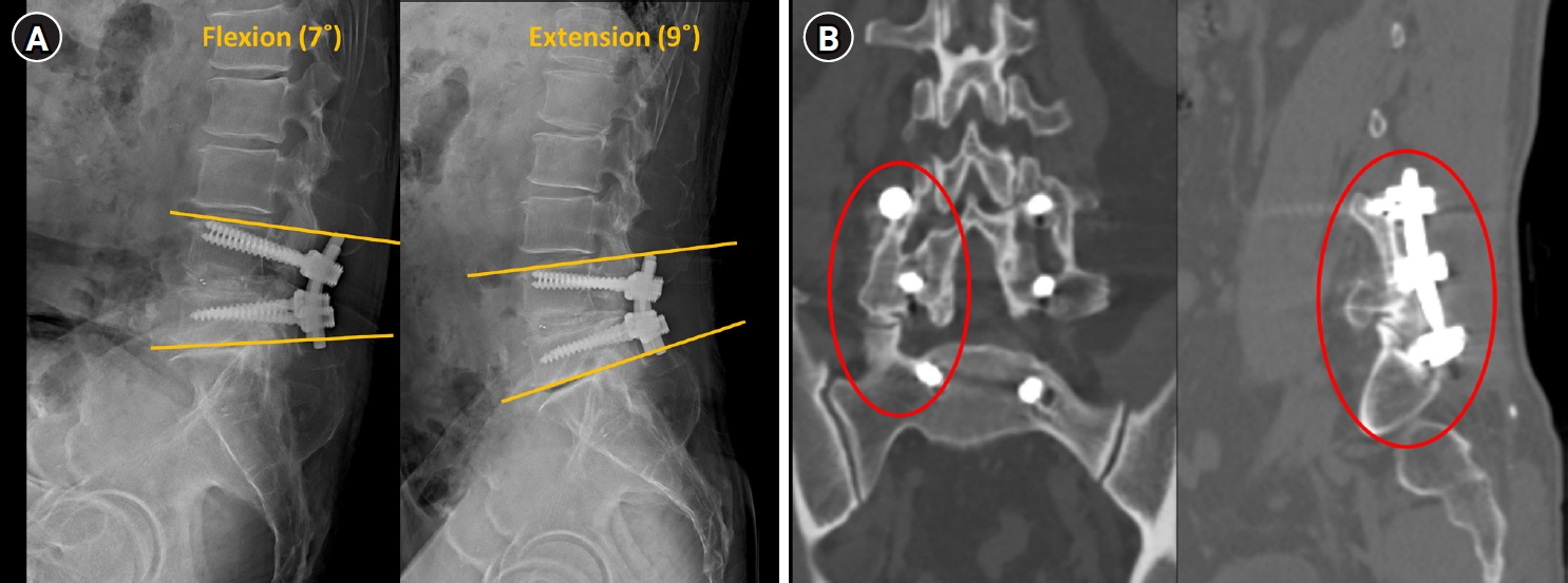
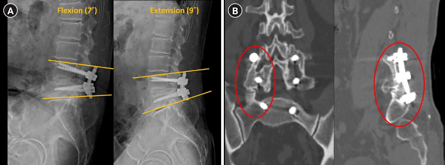
Fusion status was evaluated at the 1-year follow-up using dynamic flexion–extension X-ray and computed tomography (CT). Solid fusion was defined as ≤3° of angular motion between the upper endplate of the most superior instrumented vertebra and the lower endplate of the most inferior vertebra on dynamic X-ray (Fig. 1A).13,18-20) In eight patients who underwent postoperative CT, the presence of a continuous trabecular bone bridge across the fusion site was considered additional radiologic evidence of solid fusion (Fig. 1B).21)
2. Surgical technique
All patients underwent lumbar revision surgery through a posterior approach along the previous incision. When additional decompression or deformity correction was required, fusion extension was performed by one level above or below the original construct. Patients were placed prone on a Jackson spinal table to maintain lumbar lordosis. After midline incision and careful dissection along the prior scar, all previous screws and rods were exposed and removed. To achieve stronger fixation, screws one size larger in diameter than the original were used (CD Horizon Solera Spinal System, Medtronic, Sofamor-Danek, Memphis, TN, USA). Allograft bone chips (Maxxeus, Kettering, OH, USA) were densely packed into loosened screw holes using an impactor and mallet—approximately 15 mL per hole when enlarged. New screws were then inserted along the original trajectory, requiring firm pressure due to the compacted graft material, thereby ensuring enhanced pull-out strength. For intact tracts without loosening, simple upsizing of the screw was performed (commonly from 6.5 mm to 7.5 mm) (Supplementary Video S1). After fixation, additional decompression or pedicle subtraction osteotomy (PSO) was carried out if indicated. Rods were applied and contour-matched to restore alignment. Finally, posterolateral fusion using autologous bone from decompression and supplemental allograft was performed, and screw position and alignment were confirmed with intraoperative anteroposterior and lateral X-ray.
3. Ethics statement
This retrospective study was conducted at a tertiary referral hospital and approved by the institutional review board, which waived the requirement for informed consent (IRB No. 2024-0113).
Results
1. Demographics
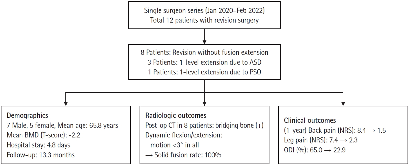
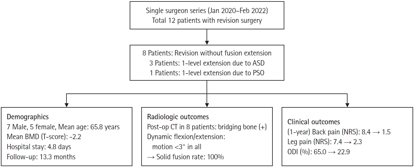
A total of 12 patients underwent revision lumbar surgery for screw loosening and nonunion (Fig. 2). The cohort included seven men and five women, with a mean age of 65.8 years, mean bone mineral density of –2.2 g/cm2, mean hospital stay of 4.8 days, and mean follow-up duration of 13.3 months. Four patients were diagnosed with osteoporosis, and all completed at least 1 year of follow-up. Eight patients underwent revision without fusion extension, three required one-level extension for ASD, and one required one-level extension with PSO for deformity correction. Detailed demographic and operative characteristics are summarized in Table 1.
2. Radiologic and clinical outcomes
At the 1-year follow-up, all patients showed ≤3° of motion on dynamic flexion–extension X-ray, confirming the absence of instability. CT was performed in eight patients and demonstrated a continuous trabecular bone bridge at the fusion site in all cases. Consequently, solid fusion was achieved in all 12 patients, corresponding to a 100% fusion rate (Table 2).
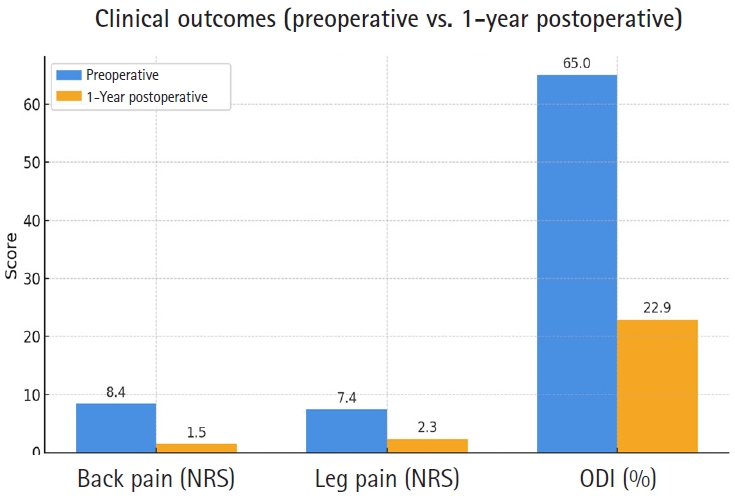
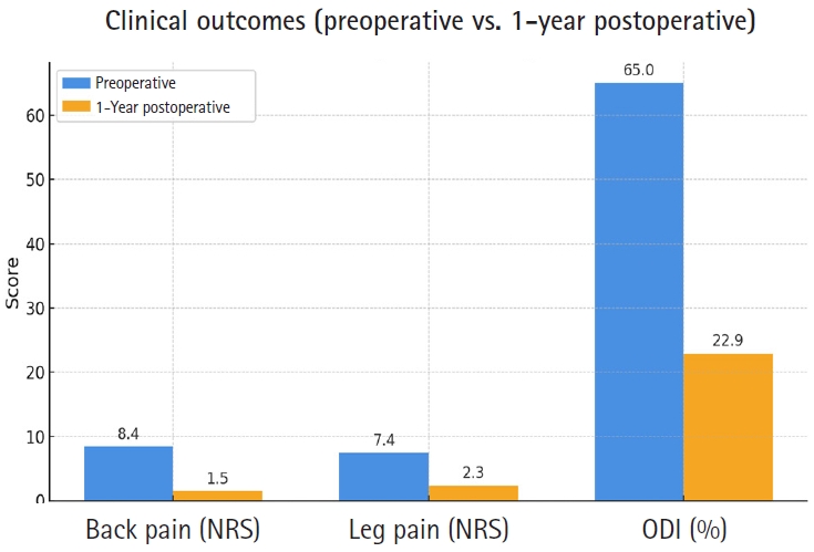
Preoperative back and leg pain scores averaged 8.4±0.9 and 7.4±1.8, respectively, which improved to 1.5±0.7 and 2.3±1.2 at 1 year, representing mean reductions of 6.9 and 5.1 points on the numerical rating scale (NRS). Functional outcomes also improved markedly, with a mean Oswestry Disability Index reduction from 65.0% to 22.9% (Table 2, Fig. 3).
3. Illustrative case
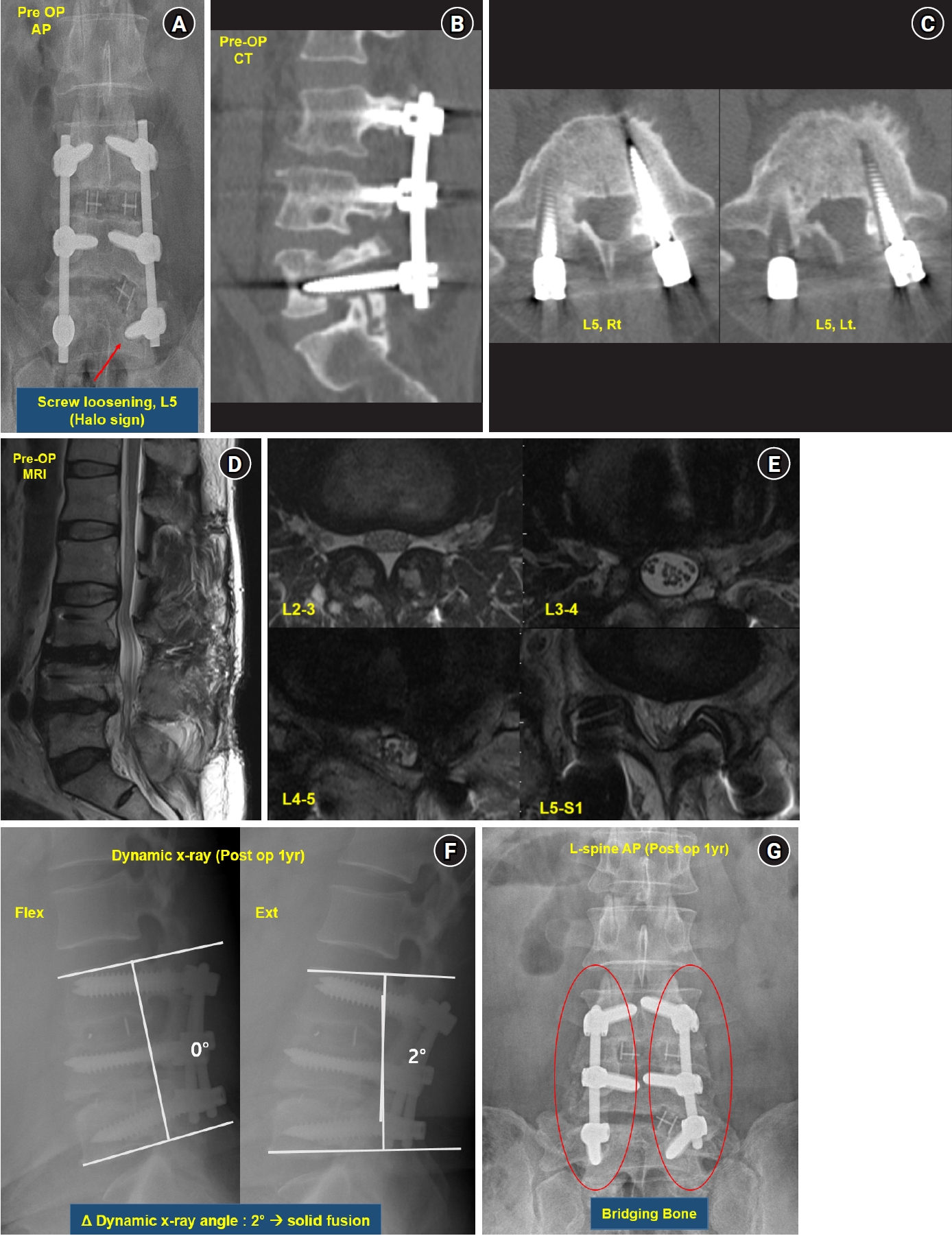
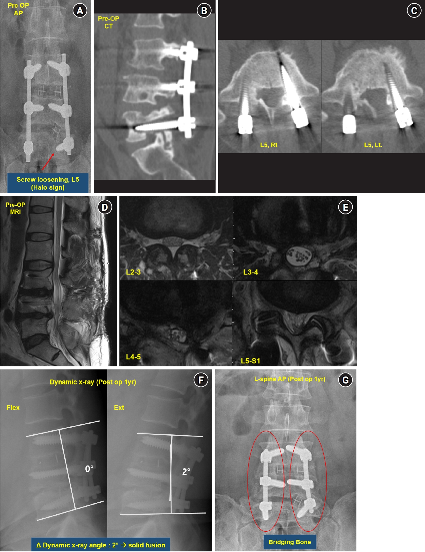
A 55-year-old man presented with persistent low back pain, bilateral leg pain, and numbness. He had previously undergone decompression at L4–5 in 2012 and posterior lumbar interbody fusion at L3–5 in 2019. Although symptoms improved temporarily, they gradually worsened, with severe back pain (NRS 9) and both leg pain (NRS 8) at presentation. Anteroposterior X-ray revealed a halo sign suggestive of L5 screw loosening, and CT confirmed bilateral L5 screw loosening with nonunion at L3–4–5 (Fig. 4A–C). Magnetic resonance imaging demonstrated preserved spinal canal dimension without significant stenosis (Fig. 4D, 4E), and neurological examination showed mild weakness (grade 4) of the left lower extremity.
Through a posterior approach, the previous instrumentation was removed. The L5 screw holes were markedly enlarged, and approximately 15 mL of allograft bone chips were densely impacted into each hole using an impactor and mallet. Larger (7.5 mm) screws were then reinserted along the original trajectory, requiring high insertional torque due to dense graft packing. An additional posterolateral fusion using autograft and allograft bone chips was also performed.
Postoperatively, the patient experienced marked relief of pain and numbness. At the 1-year follow-up, dynamic X-ray showed ≤3° of motion without instability, and anteroposterior X-ray demonstrated a distinct bony bridge at the fusion site (Fig. 4F, 4G). Back pain resolved completely (NRS 0), and leg pain improved to 3, well controlled with medication.
Discussion
This study demonstrated that short-segment fusion using allograft bone chip insertion is an effective method for treating screw loosening and nonunion after lumbar posterior fixation. Solid fusion was achieved in all 12 patients without major complications during the 1-year follow-up. To our knowledge, this is the first report focusing on the use of allograft bone chips within screw holes for short-segment lumbar revision.
Traditionally, most surgeons have managed screw loosening and nonunion by extending the fusion construct to one or more adjacent levels.14,22) However, this approach inevitably sacrifices intact motion segments and increases the lever arm, thereby raising the risk of ASD and implant failure.9,23-25) Our results suggest that short-segment revision with bone chip augmentation can achieve comparable fusion rates while avoiding the disadvantages of long-segment fusion. The allograft bone chip insertion technique is simple yet effective, allowing shorter operative times and less blood loss. In addition, minimizing the fusion length reduces bending stress on the construct, which may lower the risk of screw loosening, as reported by Marie-Hardy et al.22) These findings further support the biomechanical rationale for preserving motion segments whenever possible.
In osteoporotic patients, long-segment fusion is often associated with recurrent implant failure and proximal junctional kyphosis.26) In our series, four osteoporotic patients achieved successful fusion with short-segment fixation, demonstrating the feasibility of this technique in compromised bone. Nevertheless, adjacent segment fractures may occur due to stress concentration similar to the “hammer effect” observed after vertebroplasty.27) To mitigate these risks, our protocol includes bone-strengthening agents such as teriparatide or romosozumab and close postoperative monitoring.
In addition to fusion extension, bone cement augmentation has been considered another strategy to manage screw loosening by reinforcing fixation strength. Although this method can provide immediate mechanical stability, it carries inherent risks such as cement leakage into neural or vascular structures, potentially leading to serious complications including neurological injury or pulmonary embolism.28,29) Moreover, cement obstructs cancellous channels and impedes biological fusion. In contrast, the allograft bone chip impaction technique preserves the trabecular architecture of the host bone, enabling osteoconduction and natural bone remodeling while achieving stable fixation within the same segment.30)
Appropriate patient selection is essential when considering this technique. This technique may be most suitable for patients with localized screw loosening and nonunion, preserved overall spinal alignment, and no evidence of severe instability or progressive deformity. In such cases, reuse of the original screw trajectory with allograft bone chip insertion may provide sufficient mechanical stability and promote fusion while preserving motion segments. In contrast, patients with extensive bone loss, marked segmental instability, significant deformity, or multi-level involvement may be less suitable for this technique, as these conditions often require more robust stabilization through fusion extension. Therefore, careful preoperative assessment, including evaluation of alignment, stability, and bone quality, is crucial in determining the optimal surgical strategy for each patient.
When performing bone chip insertion, careful preoperative planning with CT is essential to confirm that the screw tract is isolated from neural structures. Because impacted bone chips can be densely compacted (up to 15 mL per hole), inadvertent communication with the spinal canal or nerve root may cause neurological injury. Intraoperative probing of each screw hole before impaction is strongly recommended.
In summary, the present study highlights a straightforward and biologically favorable approach for managing screw loosening and nonunion after lumbar fusion. By reusing existing screw trajectories and applying allograft bone chip impaction, surgeons can achieve stable fixation while minimizing surgical morbidity and preserving motion segments. This technique may serve as a practical alternative to long-segment fusion, particularly in elderly or osteoporotic patients in whom extensive instrumentation poses greater mechanical and physiological burdens.
This study has several limitations. Its retrospective design, small sample size, and single-surgeon experience may introduce selection bias and limit the generalizability of the findings. In addition, the absence of a control group precludes direct comparison with conventional revision strategies. Although fusion assessment was primarily based on flexion–extension radiographs, postoperative CT scans were performed in only a subset of patients (n=8), allowing more precise confirmation of trabecular bone bridging. However, incomplete CT evaluation across all patients may have led to underestimation or overestimation of the true fusion rate. Furthermore, the relatively short follow-up period of 1 year may be insufficient to capture delayed implant failure or ASD. Nevertheless, the consistent fusion outcomes observed in this pilot series are encouraging. Future prospective studies with larger cohorts, inclusion of appropriate control groups, standardized CT-based fusion assessment for all patients, and longer follow-up are warranted to validate the long-term efficacy of this technique.
Short-segment fusion using an allograft bone chip insertion technique appears to be a practical and biologically favorable revision option for managing screw loosening and nonunion in selected patients. It may achieve solid fusion without extending the fusion level, thereby potentially reducing surgical morbidity and preserving motion segments. Although this approach cannot replace all fusion extension procedures, it can help avoid fusion extension in selected cases and serve as a practical alternative to long-segment fusion. Larger prospective studies with standardized fusion assessment and longer follow-up are needed to confirm its broader applicability.
Revision surgery for screw loosening and nonunion using the allograft bone chip insertion technique. After exposure through the previous incision, loosened screws were removed, and the enlarged tracts were densely packed with approximately 15 mL of allograft bone chips using an impactor and mallet. New screws with a one-size larger diameter were inserted along the same trajectory, requiring high insertional torque due to the compacted graft material. This video demonstrates the key steps of bone-chip impaction, tapping, and reinsertion, highlighting a simple yet effective method for achieving solid fusion without extending the fusion level.
The authors have no conflicts of interest to declare.
Funding
None.
Acknowledgments
None.
Fig. 1.
Radiologic evaluation of fusion stability. (A) Lumbar flexion (7°) and extension (9°) lateral X-ray used to assess dynamic motion between the upper and lower instrumented vertebrae. (B) Postoperative computed tomography scans showing continuous trabecular bone bridging across the fusion site (red circles), confirming solid fusion.
Fig. 2.
Flowchart summarizing study enrollment and outcomes. Twelve patients underwent revision surgery for screw loosening and nonunion (eight without fusion extension, three with one-level extension for adjacent segmental disease (ASD), and one with pedicle subtraction osteotomy [PSO]). Radiologic and clinical results are summarized, showing 100% fusion rate and significant improvement in back/leg pain and Oswestry Disability Index (ODI). BMD: bone mineral density, CT: computed tomography, NRS: numerical rating scale.
Fig. 3.
Comparison of preoperative and 1-year postoperative clinical outcomes. Mean back pain (numerical rating scale [NRS]) improved from 8.4 to 1.5, leg pain (NRS) from 7.4 to 2.3, and Oswestry Disability Index (ODI) from 65.0% to 22.9%, indicating marked pain relief and functional recovery.
Fig. 4.
Illustrative case of revision surgery for screw loosening and nonunion. (A) Preoperative X-ray showing L5 screw loosening (halo sign). (B) Preoperative sagittal computed tomography (CT) confirming bilateral L5 screw loosening. (C) Axial CT at L5 level demonstrating enlarged screw tracts bilaterally. (D) Sagittal T2-weighted magnetic resonance imaging (MRI) showing preserved spinal canal dimension without significant stenosis. (E) Axial MRI at L3–4 and L4–5 levels showing no significant stenosis. (F) One-year postoperative dynamic X-ray showing ≤2° of motion, consistent with solid fusion. (G) One-year postoperative X-ray demonstrating continuous bridging bone formation along the fusion site (red circles).
Table 1.
Demographics and characteristics of 12 patients
Patient No.
Age (years)
Sex
BMD
Diagnosis
Previous fusion level
Operative level
Hospital stay (days)
Follow-up period (months)
1
51
M
–1.5
Screw loosening (L5)
L3-5
L3–5
4
13
2
55
M
–1.8
Screw loosening (S1)+Rod fracture
L4–S1
L4–S1
5
14
3
64
M
–2.0
Screw loosening (L2, 3)
L2–S1
L2–S1
5
12
4
55
M
–1.6
Screw loosening (L5)
L3–5
L3–5
4
13
5
66
M
–2.3
Screw loosening (L5, S1)
L5–S1
L5–S1
5
15
6
71
M
–2.6
Screw loosening (S1)
L3–S1
L3–S1
5
14
7
83
F
–3.1
Screw loosening (S1)
L5–S1
L5–S1
6
12
8
80
F
–2.8
Screw loosening (L5)
L4–5
L4–5
5
13
9
71
F
–2.1
Screw loosening (L4)+ASD
L3–4
L3–4–5
4
15
10
59
F
–1.7
Screw loosening (L3)+ASD
L3–S1
L2–S1
4
12
11
67
M
–2.0
Screw loosening (L5, S1)+ASD
L4–S1
L3–S1
5
13
12
67
F
–2.5
Screw loosening (L3)+Flatback syndrome
L3–5
L3–S1 (L4 PSO)
6
14
BMD: bone mineral density, ASD: adjacent segment degeneration, PSO: pedicle subtraction osteotomy.
Table 2.
Postoperative 1-year clinical and radiological outcomes of 12 patients
a)Δ Dynamic x-ray angle, differences in flexion/extension X-ray of the upper endplate of the vertebral body at the highest level of the surgery and the lower endplate of the vertebral body at the lowest level.
b)“+” indicates that postoperative CT was performed and bone bridging was confirmed; “-” indicates that CT was not performed (bridging not assessed).
References
1. Liljenqvist U, Hackenberg L, Link T, Halm H. Pullout strength of pedicle screws versus pedicle and laminar hooks in the thoracic spine. Acta Orthop Belg 2001;67:157-63.
2. Amirouche F, Solitro GF, Magnan BP. Stability and spine pedicle screws fixation strength: a comparative study of bone density and insertion angle. Spine Deform 2016;4:261-7.
3. Esses SI, Sachs BL, Dreyzin V. Complications associated with the technique of pedicle screw fixation: a selected survey of ABS members. Spine (Phila Pa 1976) 1993;18:2231-8.
5. Sanden B, Olerud C, Petren-Mallmin M, Johansson C, Larsson S. The significance of radiolucent zones surrounding pedicle screws: definition of screw loosening in spinal instrumentation. J Bone Joint Surg Br 2004;86:457-61.
6. DeWald CJ, Stanley T. Instrumentation-related complications of multilevel fusions for adult spinal deformity patients over age 65: surgical considerations and treatment options in patients with poor bone quality. Spine (Phila Pa 1976) 2006;31(19 Suppl):S144-51.
7. Khalid SI, Nunna RS, Maasarani S, et al. Association of osteopenia and osteoporosis with higher rates of pseudarthrosis and revision surgery in adult patients undergoing single-level lumbar fusion. Neurosurg Focus 2020;49:E6.
8. Kim HJ, Kim SG, Lee HM, et al. Risk factors associated with the halo phenomenon after lumbar fusion surgery and its clinical significance. Asian Spine J 2008;2:22-6.
9. Yuan L, Zhang X, Zeng Y, Chen Z, Li W. Incidence, risk, and outcome of pedicle screw loosening in degenerative lumbar scoliosis patients undergoing long-segment fusion. Global Spine J 2023;13:1064-71.
10. Jansen JU, Zengerle L, Hackenbroch C, Dreyhaupt J, Tao Y, Wilke HJ. Prediction of screw loosening by measuring the insertion torque in non-osteoporotic patients: an in vitro study. BMC Musculoskelet Disord 2025;26:415.
15. El Saman A, Meier S, Sander A, Kelm A, Marzi I, Laurer H. Reduced loosening rate and loss of correction following posterior stabilization with or without PMMA augmentation of pedicle screws in vertebral fractures in the elderly. Eur J Trauma Emerg Surg 2013;39:455-60.
17. Ebrahimkhani M, Arjmand N, Shirazi-Adl A. Biomechanical effects of lumbar fusion surgery on adjacent segments using musculoskeletal models of the intact, degenerated and fused spine. Sci Rep 2021;11:17892.
18. Bono CM, Bawa M, White KK, et al. Residual motion on flexion-extension radiographs after simulated lumbar arthrodesis in human cadavers. J Spinal Disord Tech 2008;21:364-71.
19. Frymoyer JW, Hanley EN Jr, Howe J, Kuhlmann D, Matteri RE. A comparison of radiographic findings in fusion and nonfusion patients ten or more years following lumbar disc surgery. Spine (Phila Pa 1976) 1979;4:435-40.
20. Kuslich SD, Ulstrom CL, Griffith SL, Ahern JW, Dowdle JD. The Bagby and Kuslich method of lumbar interbody fusion: history, techniques, and 2-year follow-up results of a United States prospective, multicenter trial. Spine (Phila Pa 1976) 1998;23:1267-78.
21. Park S, Jeong YH, Ha BJ, et al. Fusion rate of Escherichia coli-derived recombinant human bone morphogenetic protein-2 compared with local bone autograft in posterior lumbar interbody fusion for degenerative lumbar disorders. Spine J 2023;23:1877-85.
22. Marie-Hardy L, Pascal-Moussellard H, Barnaba A, Bonaccorsi R, Scemama C. Screw loosening in posterior spine fusion: prevalence and risk factors. Global Spine J 2020;10:598-602.
23. Lee SH, Lee S, Jang SW, et al. Unilateral pediculectomy and reduction with short-segment pedicle screw fixation for thoracolumbar burst fracture: a case series. World Neurosurg 2024;183:e116-26.
24. Seo DK, Kim CH, Jung SK, Kim MK, Choi SJ, Park JH. Analysis of the risk factors for unfavorable radiologic outcomes after fusion surgery in thoracolumbar burst fracture: what amount of postoperative thoracolumbar kyphosis correction is reasonable? J Korean Neurosurg Soc 2019;62:96-105.
25. Shin HK, Kim M, Lee S, et al. Surgical strategy for metastatic spinal tumor patients with surgically challenging situation. Medicine (Baltimore) 2022;101:e29560.
26. Kim DK, Kim JY, Kim DY, Rhim SC, Yoon SH. Risk factors of proximal junctional kyphosis after multilevel fusion surgery: more than 2 years follow-up data. J Korean Neurosurg Soc 2017;60:174-80.
27. Yoo CM, Park KB, Hwang SH, Kang DH, Jung JM, Park IS. The analysis of patterns and risk factors of newly developed vertebral compression fractures after percutaneous vertebroplasty. J Korean Neurosurg Soc 2012;52:339-45.
28. Kang SH, Kim KT, Park SW, Kim YB. A case of pedicle screw loosening treated by modified transpedicular screw augmentation with polymethylmethacrylate. J Korean Neurosurg Soc 2011;49:75-8.
29. Wu ZX, Gong FT, Liu L, et al. A comparative study on screw loosening in osteoporotic lumbar spine fusion between expandable and conventional pedicle screws. Arch Orthop Trauma Surg 2012;132:471-6.
30. Kim H, Lee CK, Yeom JS, Lee JH, Lee KH, Chang BS. The efficacy of porous hydroxyapatite bone chip as an extender of local bone graft in posterior lumbar interbody fusion. Eur Spine J 2012;21:1324-30.
Should Lumbar Spinal Fixation Levels Be Extended in Case of Screw Loosening and Nonunion?: Allograft Bone Chip Could Avoid Fusion Extension
Fig. 1. Radiologic evaluation of fusion stability. (A) Lumbar flexion (7°) and extension (9°) lateral X-ray used to assess dynamic motion between the upper and lower instrumented vertebrae. (B) Postoperative computed tomography scans showing continuous trabecular bone bridging across the fusion site (red circles), confirming solid fusion.
Fig. 2. Flowchart summarizing study enrollment and outcomes. Twelve patients underwent revision surgery for screw loosening and nonunion (eight without fusion extension, three with one-level extension for adjacent segmental disease (ASD), and one with pedicle subtraction osteotomy [PSO]). Radiologic and clinical results are summarized, showing 100% fusion rate and significant improvement in back/leg pain and Oswestry Disability Index (ODI). BMD: bone mineral density, CT: computed tomography, NRS: numerical rating scale.
Fig. 3. Comparison of preoperative and 1-year postoperative clinical outcomes. Mean back pain (numerical rating scale [NRS]) improved from 8.4 to 1.5, leg pain (NRS) from 7.4 to 2.3, and Oswestry Disability Index (ODI) from 65.0% to 22.9%, indicating marked pain relief and functional recovery.
Fig. 4. Illustrative case of revision surgery for screw loosening and nonunion. (A) Preoperative X-ray showing L5 screw loosening (halo sign). (B) Preoperative sagittal computed tomography (CT) confirming bilateral L5 screw loosening. (C) Axial CT at L5 level demonstrating enlarged screw tracts bilaterally. (D) Sagittal T2-weighted magnetic resonance imaging (MRI) showing preserved spinal canal dimension without significant stenosis. (E) Axial MRI at L3–4 and L4–5 levels showing no significant stenosis. (F) One-year postoperative dynamic X-ray showing ≤2° of motion, consistent with solid fusion. (G) One-year postoperative X-ray demonstrating continuous bridging bone formation along the fusion site (red circles).
Fig. 1.
Fig. 2.
Fig. 3.
Fig. 4.
Should Lumbar Spinal Fixation Levels Be Extended in Case of Screw Loosening and Nonunion?: Allograft Bone Chip Could Avoid Fusion Extension
Patient No.
Age (years)
Sex
BMD
Diagnosis
Previous fusion level
Operative level
Hospital stay (days)
Follow-up period (months)
1
51
M
–1.5
Screw loosening (L5)
L3-5
L3–5
4
13
2
55
M
–1.8
Screw loosening (S1)+Rod fracture
L4–S1
L4–S1
5
14
3
64
M
–2.0
Screw loosening (L2, 3)
L2–S1
L2–S1
5
12
4
55
M
–1.6
Screw loosening (L5)
L3–5
L3–5
4
13
5
66
M
–2.3
Screw loosening (L5, S1)
L5–S1
L5–S1
5
15
6
71
M
–2.6
Screw loosening (S1)
L3–S1
L3–S1
5
14
7
83
F
–3.1
Screw loosening (S1)
L5–S1
L5–S1
6
12
8
80
F
–2.8
Screw loosening (L5)
L4–5
L4–5
5
13
9
71
F
–2.1
Screw loosening (L4)+ASD
L3–4
L3–4–5
4
15
10
59
F
–1.7
Screw loosening (L3)+ASD
L3–S1
L2–S1
4
12
11
67
M
–2.0
Screw loosening (L5, S1)+ASD
L4–S1
L3–S1
5
13
12
67
F
–2.5
Screw loosening (L3)+Flatback syndrome
L3–5
L3–S1 (L4 PSO)
6
14
Patient No.
Δ Dynamic X-ray anglea)
Bone bridging on CTb)
Back pain (NRS)
Leg pain (NRS)
ODI (%)
Pre
1 Year
Δ(Pre–1 year)
Pre
1 Year
Δ(Pre–1 year)
Pre
1 Year
Δ(Pre–1 year)
1
2
+
9
0
9
8
3
5
71
18
53
2
1
+
5
2
3
5
2
3
45
25
20
3
0
+
8
4
4
4
4
0
56
39
17
4
2
–
10
4
6
7
4
3
73
40
33
5
2
–
7
4
3
7
4
3
59
38
21
6
1
+
10
0
10
9
1
8
78
13
65
7
0
+
7
1
6
7
2
5
59
22
37
8
1
+
7
0
7
10
2
8
67
15
52
9
0
+
10
1
9
8
2
6
55
14
41
10
2
–
9
0
9
9
1
8
73
13
60
11
0
–
9
2
7
9
2
7
74
25
49
12
2
+
10
0
10
6
1
5
70
13
57
Table 1. Demographics and characteristics of 12 patients
BMD: bone mineral density, ASD: adjacent segment degeneration, PSO: pedicle subtraction osteotomy.
Table 2. Postoperative 1-year clinical and radiological outcomes of 12 patients
Δ Dynamic x-ray angle, differences in flexion/extension X-ray of the upper endplate of the vertebral body at the highest level of the surgery and the lower endplate of the vertebral body at the lowest level.
“+” indicates that postoperative CT was performed and bone bridging was confirmed; “-” indicates that CT was not performed (bridging not assessed).


 , Jin Hoon Park, M.D., Ph.D.1)