1)Department of Neurological Surgery, Oregon Health & Science University, Portland, OR, USA
2)Department of Orthopaedics and Rehabilitation, Oregon Health & Science University, Portland, OR, USA
Corresponding author: Christina Gerges Castro, M.D. Department of Neurological Surgery, Oregon Health & Science University, 3181 SW Sam Jackson Park Rd., Portland, OR 97239, USA TEL: +1-503-494-4314, FAX: +1-503-346-6810, E-mail: Gerges@ohsu.edu
• Received: February 27, 2026 • Revised: April 29, 2026 • Accepted: May 15, 2026
This is an open access article distributed under the terms of the Creative Commons Attribution Non-Commercial License (https://creativecommons.org/licenses/by-nc/4.0/) which permits unrestricted non-commercial use, distribution, and reproduction in any medium, provided the original work is properly cited.
Traumatic lumbar spondyloptosis is a rare entity associated with high-velocity mechanisms and is the most severe form of lumbar spondylolisthesis. Operative management is often required; however, the relative merits of reduction versus in situ fusion remain debated, largely owing to the technical difficulty of attaining satisfactory fracture reduction. In this report, we describe external femoral traction as a novel technique for closed reduction of traumatic lumbar spondyloptosis. A 27-year-old man presented after a tree he was cutting fell on him and was found to have T3–7 AO Spine (AOS) A1 fracture, L3 AOS B2 fracture, and L5 AOS C fracture. Neurologic exam was consistent with multilevel nerve root injury. Definitive treatment included bilateral femoral traction, open reduction, and combined anterior/posterior fixation. A multidisciplinary team including orthopedic surgery, plastic surgery, vascular surgery, and neurosurgery were involved. Complete reduction was obtained, and the patient experienced near-complete resolution of neurologic symptoms. This technique offers a unique solution to the challenge of traumatic lumbar spondyloptosis. Further study and follow-up are needed to confirm the utility and durability of this technique and the cranial extent of injury for which this technique might be applied.
Traumatic lumbar spondyloptosis is a rare entity associated with high velocity mechanisms and is the most severe form of lumbar spondylolisthesis (Meyerding Grade V).1-3) Patients often present with significant polytrauma and may experience back pain, spinal deformity, or neurologic deficits of variable severity. Operative management is required for reduction and stabilization. The primary operative technique previously described in the literature included open reduction with osteotomies and internal fixation.1,4-7) A single 2013 study reported closed reduction of traumatic spondyloptosis using lumbar hyperextension and full torso longitudinal traction followed immediately by open internal fixation.3) To date, no other reports regarding external reduction of traumatic lumbar spondyloptosis exist. In this report, we describe an external femoral traction as a novel technique for closed reduction of traumatic lumbar spondyloptosis.
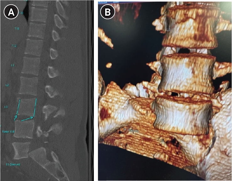
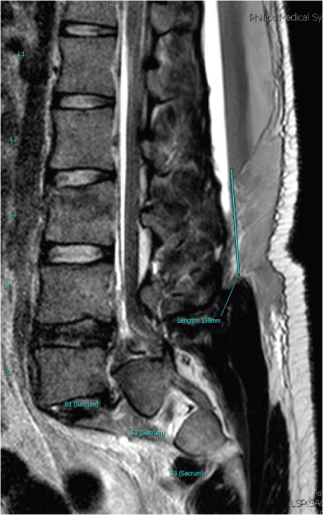
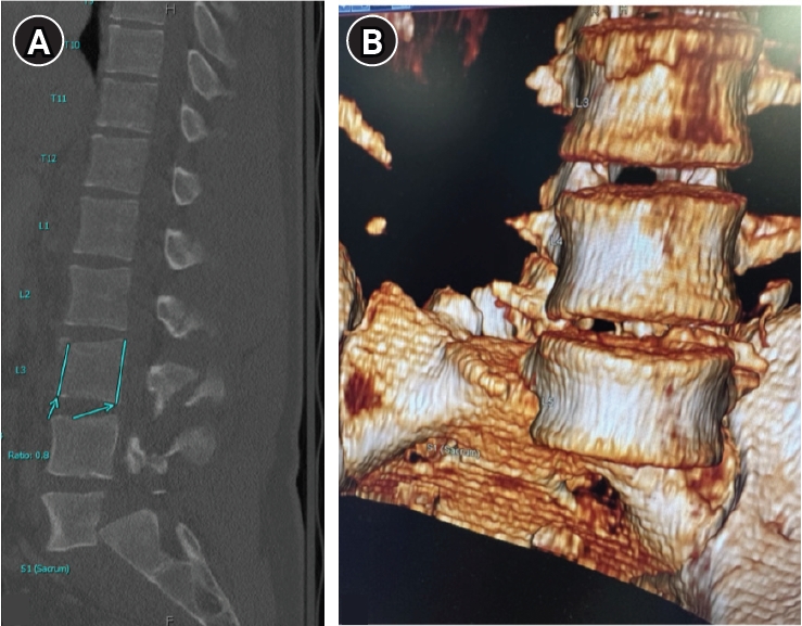
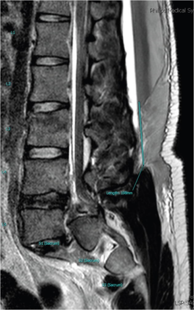
A 27-year-old man presented to the emergency room as a trauma activation after a tree he was cutting fell on him. On initial neurosurgical evaluation, he was found to have bilateral hip flexion weakness (motor score 3/5 bilaterally), minimal movement in ankle dorsiflexion and plantar flexion (motor score 1/5 bilaterally), and diminished sensation to light touch in bilateral S1 dermatomes. Foley catheterization was required for urinary retention. Rectal tone was spared. Computed tomography (CT) of the spine demonstrated T3–7 AO Spine (AOS)8) A1 fracture, L3 AOS B2 fracture, and L5 AOS C fracture (Fig. 1). Vascular imaging of the abdomen was obtained due to significant risk of large vessel injury with this pathology, although no vascular injury was noted. Magnetic resonance imaging (MRI) of the spine was subsequently obtained (Fig. 2). A waiver of informed consent was granted due to the absence of patient identifiers.
Technical Note
1. Initial approach
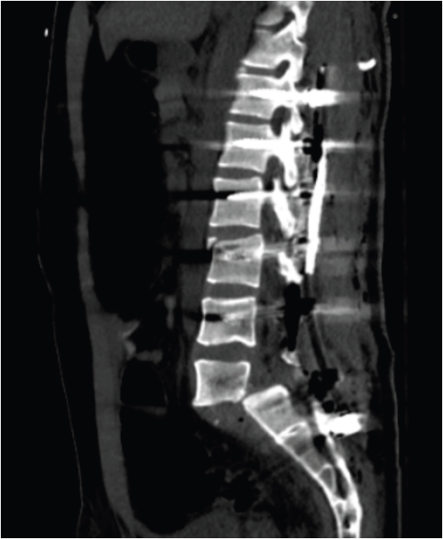
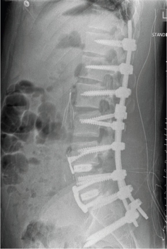
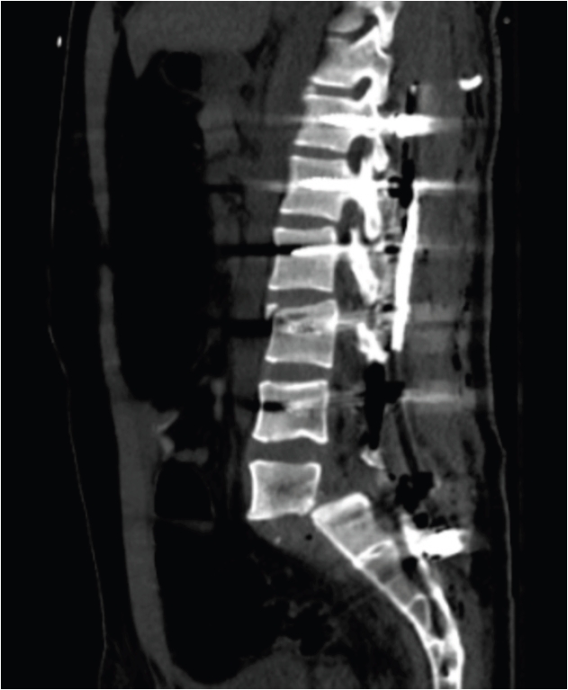
An initial attempt at standard open reduction was performed, including facetectomies, discectomies, and the use of reduction towers with posterior segmental instrumentation extending from T12 to the pelvis. This upper instrumented vertebra was selected to bypass the thoracolumbar junction and provide a stable proximal anchor in the setting of a high-grade L5 AOS C-type fracture with a long lever arm and multilevel injury. Unfortunately, the reduction could not be maintained. Traumatic durotomies sustained at the time of injury were repaired primarily, followed by a muscle graft and dural sealant. Postoperative CT imaging demonstrated persistent L5–S1 spondyloptosis, with the L5 vertebral body remaining ventral to the sacrum (Fig. 3). The patient was subsequently returned to the operating room for a reduction attempt using femoral traction.
2. Distal femoral traction
The patient was positioned supine on the operating room table and was sterilely prepared and draped. A sterile 2.0-mm Kirschner wire was selected from the skeletal traction tray and advanced percutaneously through the distal femoral metaphysis from lateral to medial using a motorized wire driver. Care was taken to ensure a tension-free skin exit site. The sharp ends were protected, the Kirschner traction bow was attached to the wire, and 40 pounds of longitudinal traction was placed on each extremity using rope, sandbags, and a pulley system.
3. Revision fusion and spondyloptosis reduction
The previous incision was opened. The previous rods were temporarily removed. The L5 level was identified, and the facet and disrupted pedicle remnant were removed with Leksell rongeurs with care to protect the traversing and exiting roots. A Cobb elevator was inserted to distract across the disc space; however, reduction was achieved almost entirely through the application of longitudinal traction rather than manual manipulation. A temporary rod was placed from L4–S1 on the right. An appropriately contoured rod was positioned on the left side to span the entirety of the construct, and set caps were applied and sequentially tightened to preserve the achieved reduction. The temporary right-sided rod was subsequently removed and replaced with an appropriately sized and contoured rod, after which the set caps were placed and fully tightened. Upon completion of posterior fixation, the 40-pound sandbags were removed from the traction pulley system.
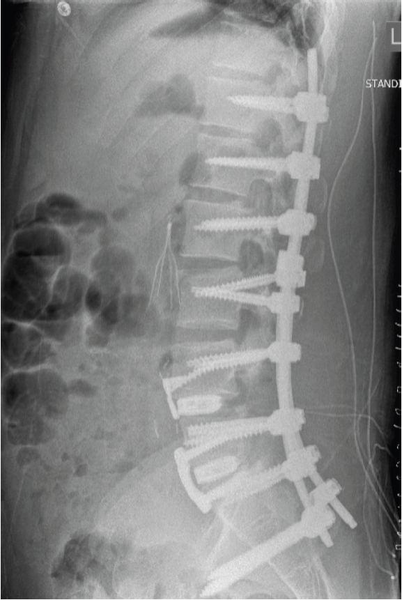
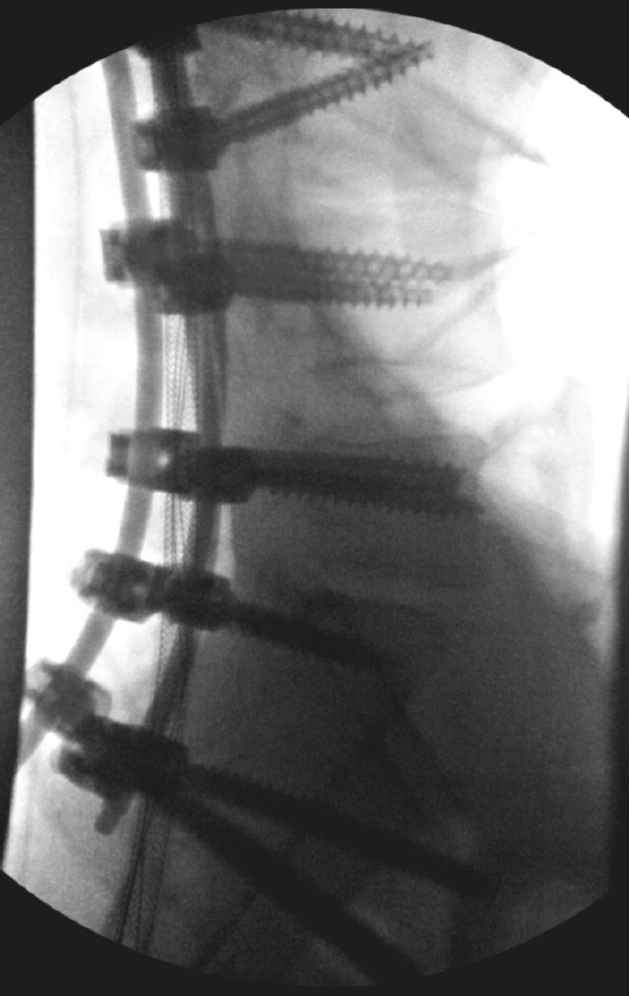
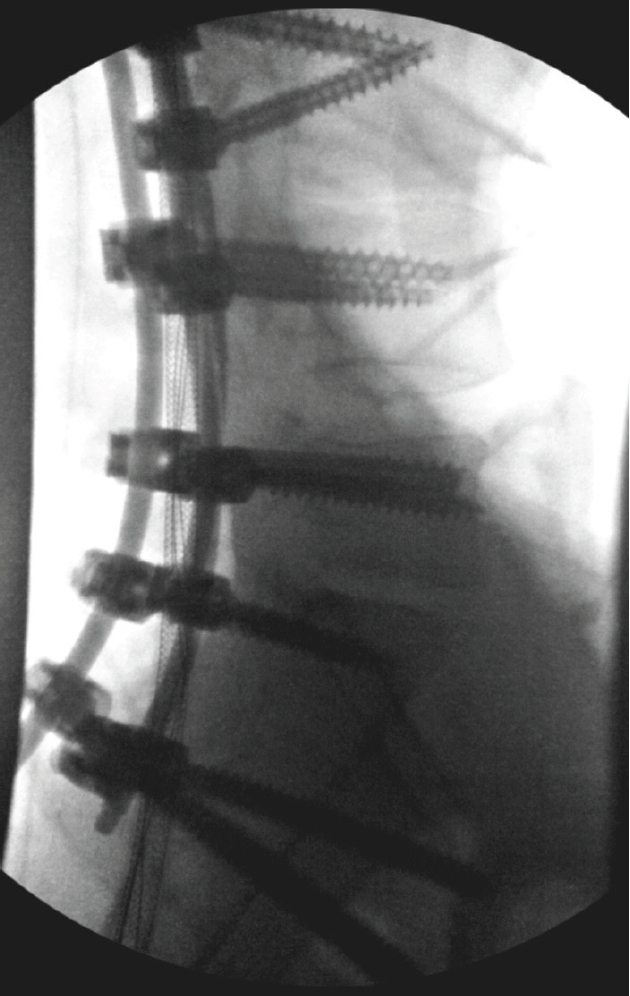
A lumbar drain was placed at the L3–4 interspace under direct visualization and secured. Fusion allograft and autograft material were placed over the lamina, facets, and transverse processes of the instrumented levels. Complex spine closure was performed by a plastic surgeon. Hardware disposition following this portion of the procedure is available in Fig. 4.
4. Anterior lumbar interbody fusion
Following closure, the patient was rotisserie-flipped for an anterior approach. A vascular surgeon exposed the anterior lumbar spine from L4–S1. Discectomy was completed at L5/S1 using standard techniques. A trial was sized. Using fluoroscopic guidance, an anterior lumbar interbody fusion cage was placed and secured. This process was repeated at L4–5 to augment the adjacent segment, improve load sharing, and enhance construct stability across the lumbosacral junction. The anterior incision was then closed. Final hardware disposition is available in Fig. 5.
5. Removal of traction pin
The pin was cut with the skin on one end. It was cleaned with chlorhexidine and pulled through the contralateral end carefully.
6. Postoperative course
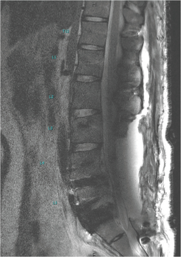
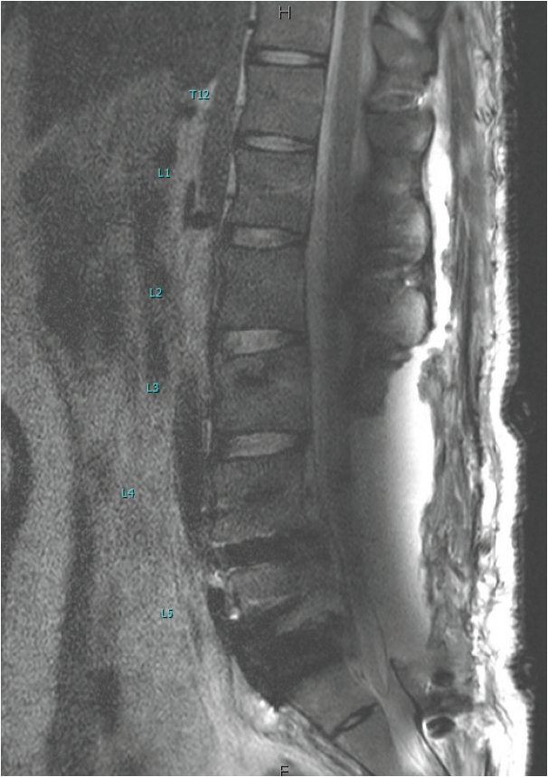
A pseudomeningocele was appreciated on MRI (Fig. 6). The lumbar drain was weaned and eventually removed. Following an uneventful postoperative course, the patient was discharged to an inpatient rehabilitation facility with residual bilateral foot drop and urinary retention requiring regular straight catheterization. At the 6-week postoperative visit, all urinary symptoms had resolved. By the 1-year postoperative visit, his foot drop had resolved bilaterally. He was ambulatory without ankle-foot orthotics with no bladder or bowel symptoms. His only residual symptoms currently are minimal back pain and bilateral foot numbness.
Discussion
Lumbar spondyloptosis represents one of the most extreme forms of translational spinal instability. Given the scarcity of high-quality evidence, there remains no consensus for optimal surgical management. Reduction is technically demanding and includes restoration of alignment, decompression of neural elements, and fixation across a severely destabilized segment. In traumatic cases of spondyloptosis, early reduction and fusion have been associated with significant improvements in patient-reported pain, disability scores, and radiographic alignment. Xu et al.4) and Zhou et al.9) reported complete reduction and fusion in traumatic lumbar spondyloptosis, with patients achieving pain relief, neurologic improvement, restored alignment, and solid bony fusion.
In situ fusion has been proposed as an alternative when in chronic spondyloptosis or when reduction is not feasible or deemed unsafe. No reports of in situ fusion with or without decompression for acute traumatic spondyloptosis were identified. Wangtaphan et al.10) and Emel et al.11) described cases of delayed presentation of traumatic lumbosacral spondyloptosis wherein in situ fusion was performed, resulting in improved back pain and bony fusion at the level of injury. While in situ fusion avoids risks associated with reduction maneuvers, it has several drawbacks, including persistent deformity and potential for chronic pain related to malalignment. Additionally, fusion without reduction may compromise the ability to achieve robust biomechanical fixation, increasing the risk of pseudarthrosis, although this has not been reported for this pathology.
In this technical note, we describe the novel application of bilateral femoral traction as an adjunctive method for achieving controlled reduction of lumbar spondyloptosis prior to posterior fixation. The biomechanical rationale for femoral traction lies in the ability to generate longitudinal distraction across the lumbosacral junction, counteracting the anterior shearing vector that is characteristic of the pathology. By applying traction through bilateral femoral pins, realignment may be achieved through ligamentotaxis and soft-tissue tensioning. This can decrease the need for forceful intraoperative manipulation, restore relative anatomical orientation, and facilitate subsequent instrumentation.
While skeletal traction has been described for cervical and thoracic spondyloptosis and spinopelvic dissociation, its use in the lumbosacral spine remains rarely reported.3,12-14) To our knowledge, the use of femoral traction as a reduction maneuver for this pathology has not been described in the literature.
This technique offers several practical advantages. It allows controlled, titratable traction using readily available equipment. It can be performed with the patient in the supine or prone position, depending on the planned surgical approach. It enables continuous radiographic monitoring to gauge reduction progress and tissue response. Furthermore, by restoring partial alignment prior to exposure, traction can facilitate localization, instrumentation, and reduction maneuvers during definitive fixation.
However, several precautions and limitations should be emphasized. Excessive or rapid traction may risk neurovascular injury, particularly stretch injury to the lumbosacral plexus or compromise of iliac and femoral vessels. Importantly, traction is not a substitute for definitive stabilization; rather, it is an adjunct to facilitate safer and more controlled alignment restoration.
In summary, femoral traction provides a simple and reproducible adjunct for the reduction of traumatic lumbar spondyloptosis. When applied judiciously under imaging guidance, it may enhance the safety and efficiency of subsequent open reduction and fixation. Future studies and accumulation of technical experience are needed to better define patient selection criteria, optimal traction parameters, and long-term outcomes associated with this approach.
This technique offers a unique solution to the challenge of traumatic lumbar spondyloptosis. Further study and follow-up are needed to confirm the utility and durability of this technique and the cranial extent of injury for which this technique might be applied.
The authors have no conflicts of interest to declare.
Funding
None.
Acknowledgments
None.
Fig. 1.
(A) Sagittal computed tomography (CT) image at the time of presentation demonstrating L3 AO Spine B2 fracture, and L5 spondyloptosis. (B) 3-D reconstruction of CT imaging demonstrating anterior inferior displacement of the L5 vertebral body over the sacrum.
Fig. 2.
Sagittal T2 sequence magnetic resonance imaging redemonstrating L3 AO Spine (AOS) B2 fracture and L5 AOS C fracture.
Fig. 3.
Sagittal computed tomography demonstrating initial unsuccessful attempt at open reduction.
Fig. 4.
Intraoperative X-ray taken after spondyloptosis reduction and posterior instrumentation.
Fig. 5.
Postoperative X-ray demonstrating final hardware disposition (L4/5, L5/S1 anterior lumbar interbody fusion and T12-pelvis posterior segmental fusion).
Fig. 6.
Postoperative sagittal T2 orthopedic-metal artifact reduction sequence magnetic resonance imaging demonstrating pseudomeningocele secondary to durotomies sustained at the time of injury.
References
1. Braithwaite J, Gruber J, Fakhoury J, Katsigiorigis G, Grewal K. Acute traumatic spondyloptosis: a case report. Cureus 2023;15:e36457.
3. Francis T, Steinmetz M, Moore T. Traumatic spondyloptosis of the lumbar spine: closed reduction and internal fixation. Spine (Phila Pa 1976) 2013;38:E1636-40.
5. Xu F, Tian Z, Fu C, et al. Mid-lumbar traumatic spondyloptosis without neurological deficit: a case report and literature review. Medicine (Baltimore) 2020;99:e19578.
7. Akesen B, Mutlu M, Kara K, Aydinli U. Traumatic lumbosacral spondyloptosis: a case report and review of the literature. Global Spine J 2014;4:59-62.
10. Wangtaphan W, Oo M, Paholpak P, Wang Z, Sakakibara T, Kasai Y. Traumatic lumbosacral spondyloptosis treated five months after injury occurrence: a case report. Spine (Phila Pa 1976) 2012;37:E1410-4.
11. Emel E, Ozer F, Karagoz Guzey F, Alatas I, Guzey D. Lumbosacral spondyloptosis treated by two-staged fusion in situ operation: a case report. Turk Neurosurg 2006;16:145-9.
12. Ramieri A, Domenicucci M, Cellocco P, Lenzi J, Dugoni DE, Costanzo G. Traumatic spondylolisthesis and spondyloptosis of the subaxial cervical spine without neurological deficits: closed re-alignment, surgical options and literature review. Eur Spine J 2014;23 Suppl 6:658-63.
13. Anokwute MC, House H, Huh A, Ordaz J, McVeigh L, Stockwell DW. Systematic review of traumatic thoracic spondyloptosis and presentation of a novel approach for management with quad rod construct. Oper Neurosurg 2022;23:e237-44.
14. Williams SK, Quinnan SM. Percutaneous lumbopelvic fixation for reduction and stabilization of sacral fractures with spinopelvic dissociation patterns. J Orthop Trauma 2016;30:e318-24.
External Femoral Traction for Reduction of Traumatic Lumbar Spondyloptosis: A Technical Note
Fig. 1. (A) Sagittal computed tomography (CT) image at the time of presentation demonstrating L3 AO Spine B2 fracture, and L5 spondyloptosis. (B) 3-D reconstruction of CT imaging demonstrating anterior inferior displacement of the L5 vertebral body over the sacrum.
Fig. 2. Sagittal T2 sequence magnetic resonance imaging redemonstrating L3 AO Spine (AOS) B2 fracture and L5 AOS C fracture.
Fig. 3. Sagittal computed tomography demonstrating initial unsuccessful attempt at open reduction.
Fig. 4. Intraoperative X-ray taken after spondyloptosis reduction and posterior instrumentation.
Fig. 5. Postoperative X-ray demonstrating final hardware disposition (L4/5, L5/S1 anterior lumbar interbody fusion and T12-pelvis posterior segmental fusion).
Fig. 6. Postoperative sagittal T2 orthopedic-metal artifact reduction sequence magnetic resonance imaging demonstrating pseudomeningocele secondary to durotomies sustained at the time of injury.
Fig. 1.
Fig. 2.
Fig. 3.
Fig. 4.
Fig. 5.
Fig. 6.
External Femoral Traction for Reduction of Traumatic Lumbar Spondyloptosis: A Technical Note


 , Isaac Lapite, M.D.2)