Department of Neurosurgery and Medical Research Institute, Pusan National University Hospital, Pusan National University School of Medicine, Busan, Korea
Corresponding author: Dong Hwan Kim, M.D. Department of Neurosurgery and Medical Research Institute, Pusan National University Hospital, Pusan National University School of Medicine, 179 Gudeok-ro, Seo-gu, Busan 49241, Korea TEL: +82-51-240-7257, FAX: +82-51-244-0282, E-mail: smile0402@hanmail.net
• Received: December 18, 2025 • Revised: February 24, 2026 • Accepted: March 17, 2026
This is an open access article distributed under the terms of the Creative Commons Attribution Non-Commercial License (https://creativecommons.org/licenses/by-nc/4.0/) which permits unrestricted non-commercial use, distribution, and reproduction in any medium, provided the original work is properly cited.
Chondroma is a benign cartilaginous neoplasm, rarely encountered in the lumbar spine. We report a case involving a 70-year-old female who presented with lower limb radiating pain that started four years ago. Although no pronounced muscle weakness was noted, the patient experienced progressively worsening radiating pain in the L4 dermatome. Initial evaluation, including magnetic resonance imaging (MRI) performed at another medical facility, suggested a potential lesion associated with left L4–5 lumbar disc herniation. Despite non-surgical interventions, the patient experienced limited symptomatic relief, prompting her to seek further care at our clinic. Subsequent contrast-enhanced MRI conducted at our facility revealed a mass exhibiting peripheral rim enhancement surrounding the L4 nerve root. Suspecting a neurogenic tumor, we decided to perform surgical excision of the mass. Postsurgery, histopathological analysis confirmed the presence of hyaline cartilage with lobular architecture and chondrocytes in lacunae, leading to the conclusive diagnosis of chondroma. Following the surgical procedure, the previously reported radiating pain exhibited notable improvement.
Chondroma, a benign tumor originating in cartilaginous structures, presents a distinctive challenge in medical diagnosis. Although it can potentially manifest in any bone structure, its primary occurrence is observed in the long bones of the hands and feet.1) The incidence rate is exceptionally low, making the diagnosis of chondroma in the lumbar spine particularly challenging. This challenge is exacerbated by the infrequent utilization of contrast-enhanced gadolinium imaging, especially in patients presenting with symptoms such as back pain and radiating discomfort in the lower extremities. This report describes a case of chondroma in the lumbar region of a 70-year-old female who presented with radiating pain in her lower extremities.
Case Report
A 70-year-old female with osteoporosis presented with a history of low back pain accompanied by numbness in her left lower extremities persisting for the past 4 years. The patient reported severe radiating pain localized to the left L4 sensory dermatome, exacerbated by walking. Notably, there was no observed motor weakness upon neurological examination. A magnetic resonance imaging (MRI) obtained at another medical facility approximately eight months before admission indicated a suspected far lateral disc on the left side of the lumbar 4/5 region.
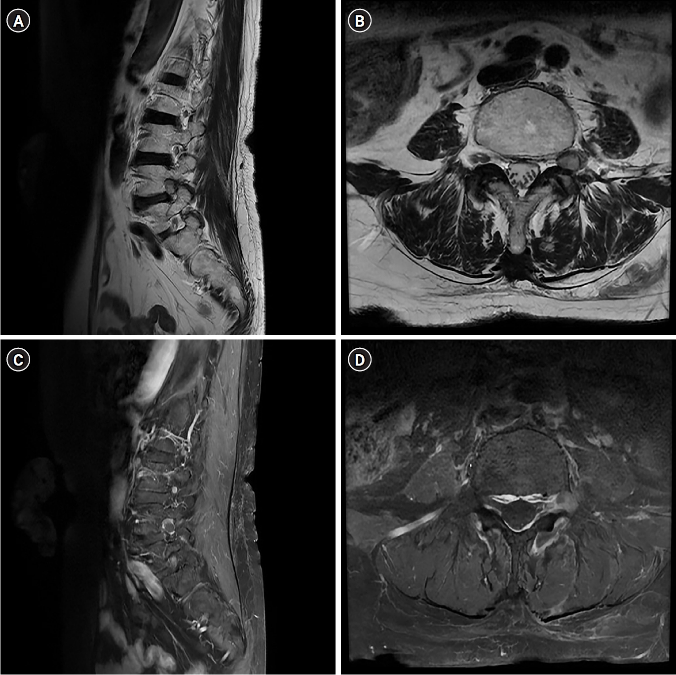
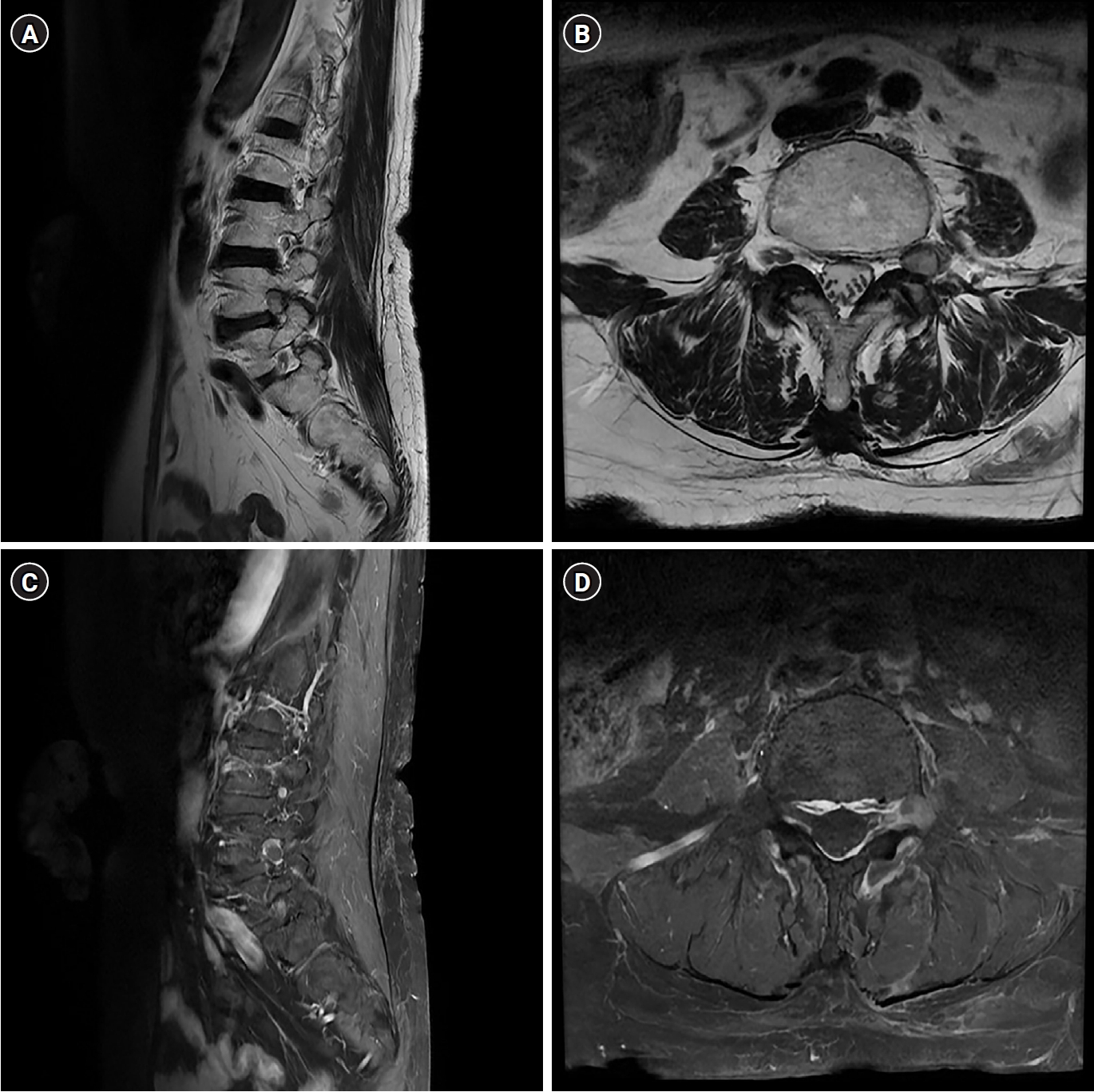
The patient underwent a left L4–5 transforaminal epidural block, which resulted in only 10 days of temporary relief. Subsequent contrast-enhanced MRI conducted at our hospital revealed a benign neurogenic tumor with cystic degeneration. The tumor demonstrated high signal intensity on T2-weighted imaging, low signal intensity on T1-weighted imaging, and peripheral enhancement on gadolinium-enhanced MRI. Measuring approximately 8 mm in size, the mass displayed a well-defined round shape and was located at the left neural foramen of the L4–5 level, causing severe compression of the left exiting L4 nerve root or potentially arising from the L4 nerve root (Fig. 1).
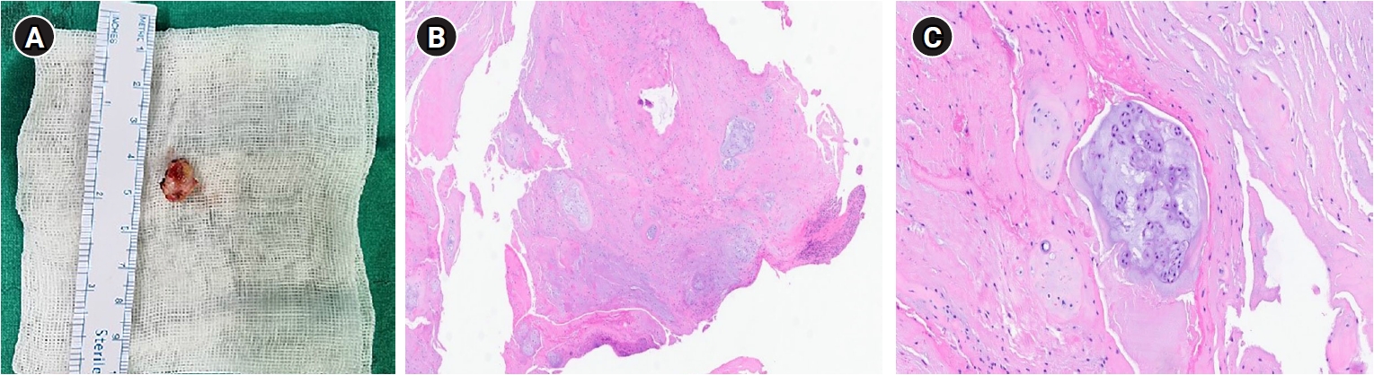
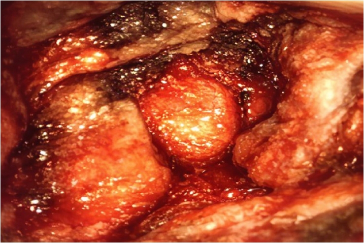
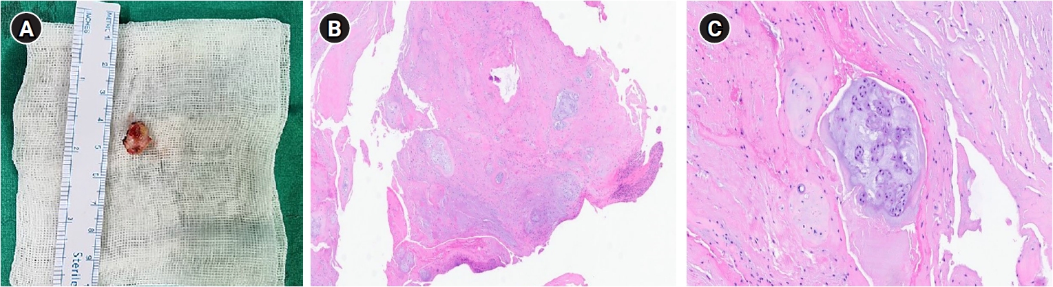
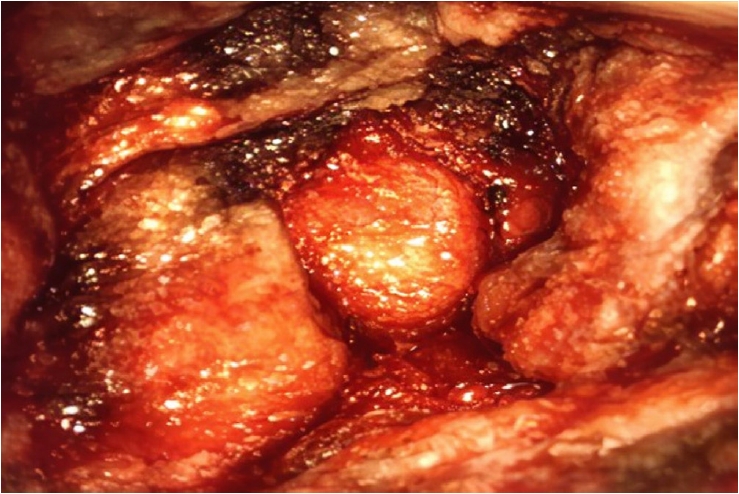
Subsequently, a surgical intervention involving L4–5 left facetectomy and complete gross resection of the extradural mass was performed, suspecting a neurogenic tumor. In the surgical field, a well-marginated yellowish mass was discovered beneath the L4 root (Fig. 2). Histopathological examination confirmed the diagnosis of chondroma (Fig. 3).
Immediately postoperation, the radiating pain in the L4 dermatome exhibited improvement, and the patient was discharged without any complications.
Discussion
Chondroma, constituting 5% of all bone tumors, is the most prevalent cartilaginous tumor.1,2) While its typical occurrence is in the long bones of the hands and feet, it can manifest in uncommon sites such as the ribs, pelvis, and spine. Notably, spinal chondromas are exceptionally rare, accounting for approximately 3% of all chondromas.1) Symptomatic chondromas within the lumbar spine are particularly scarce, with only 22 reported cases (Table 1).1-16)
Chondroma can be categorized based on their location into enchondromas (within the medullary cavity), periosteal chondromas (developing within and beneath periosteal connective tissue), and soft tissue chondromas, also referred to as synovial chondromas (found in extra-osseous and extra-synovial soft tissue regions).3,17)
Therefore, distinguishing between periosteal and soft tissue chondromas becomes challenging when a chondroma is located extradural in the spinal canal. In our case, the mass is identified as a soft tissue chondroma due to its extra-osseous location.
Depending on the tumor's location, various neurological signs and symptoms may arise. Spinal chondromas can invade the vertebral body, pedicle, lamina, and transverse/spinous processes, leading to radiculopathy, as observed in this case, or myelopathy if cord compression occurs.4) In our literature review, the neural arch was the most commonly affected location, with back pain and radiculopathy being the predominant associated symptoms in the lumbar spine.
The duration of symptoms before chondroma diagnosis varied, ranging from days to years, attributed to its slow-growing pattern and frequent misdiagnosis. The clinical characteristics, such as an acute onset of back pain with leg pain, closely mimic those of lumbar disc herniation, as demonstrated in our case. The absence of routine use of gadolinium in cases of lumbar radiculopathy contributes to the exclusion of spinal chondroma from diagnostic considerations in these patients. Even with contrast-enhanced MRI, the lesion is often initially misinterpreted as a neurogenic or dumbbell tumor extending into the foramen and extraforaminal area rather than a rare chondroma. Therefore, spinal chondroma is unlikely to be considered initially. Nonetheless, the aforementioned findings suggest the value of administering contrast to rule out this pathology in patients with lumbar disc herniation exhibiting unusual characteristics, such as inadequate response to treatment or foraminal-extraforaminal location.
Although these findings were not observed on computed tomography (CT) in the present study, in some reported cases CT has demonstrated calcification or focal bony destruction, thereby aiding in the assessment of the relationship between the tumor and adjacent osseous structures. MRI is valuable for evaluating soft tissue extension and confirming the diagnosis.18) Chondromas typically exhibit low signal intensity in T1-weighted images, high signal intensity in T2-weighted images, and peripheral enhancement with gadolinium contrast in MRI.4,19) Without contrast enhancement, differentiation from disc herniation based solely on symptoms and imaging findings can be challenging. Furthermore, even with contrast-enhanced imaging, distinguishing it from nerve sheath tumors can be difficult if the findings are non-specific, and the mass shows enhancement along the neural foramen.3,4) According to previous reports, sequestrated disc fragments may demonstrate varying degrees of peripheral rim enhancement, depending on the extent of adjacent angiogenesis and granulation tissue formation resulting from the associated inflammatory response.20) Therefore, biopsy remains indispensable for an accurate diagnosis. Histopathological findings of chondroma typically reveal chondrocytes arranged in a pseudolobular fashion and may be associated with ossified regions.5)
Despite the scarcity of reported cases and the absence of known malignant transformations, surgical resection is recommended for patients with uncontrolled pain, neurological deficit, or accelerated growth.
Spinal chondroma is an exceedingly uncommon condition characterized by a paucity of symptoms. However, when symptoms manifest, differentiating it from nerve sheath tumors and disc herniation can be challenging. Consequently, it is imperative to consider chondroma as a potential differential diagnosis, especially when contrast-enhanced MRI reveals peripheral rim enhancement, characteristic of this lesion.
The authors have no conflicts of interest to declare.
Funding
None.
Acknowledgments
This work was supported by clinical research grant in 2022.
Fig. 1.
Magnetic resonance finding of the tumor. (A) The tumor-like lesion demonstrated high signal intensity on T2-weighted imaging. (B) Low signal intensity on T1-weighted imaging. (C, D) Peripheral enhancement on T1-enhancement imaging.
Fig. 2.
In the surgical field, a well-marginated yellowish mass beneath the L4 root.
Fig. 3.
Biopsy finding. (A) Hyaline cartilage with lobular architecture, separated by fibrous bands (foci of calcification). (B, C) Histopathological examination with hematoxylin and eosin (H&E) staining revealed chondrocytes within lacunae showing uniform nuclei without atypia, with focal binucleation (B: ×40, C: ×200).
Table 1.
Reported cases of symptomatic lumbar spine chondromas
3. Cho HJ, Shim JC, Kim HK, et al. A soft tissue chondroma originating from the dura mater of the lumbar spinal canal and it mimicked a nerve sheath tumor: a case report with the MR imaging. J Korean Soc Radiol 2009;60:221-5.
10. Erten SF, Kocak A, Mizrak B, Kutlu R, Colak A. An end-plate chondroma mimicking calcified lumbar disc herniation: a case report and review of the literature. Neurosurg Rev 1999;22:145-8.
13. Cetinkal A, Guven G, Topuz AK, Colak A, Demircan MN, Haholu A. Lumbar spinal chondroma presenting with radiculopathy: case report. Turk Neurosurg 2008;18:397-9.
14. Pace J, Lozen AM, Wang MC, Cochran EJ. Extradural chondroma presenting as lumbar mass with compressive neuropathy. J Craniovertebr Junction Spine 2014;5:131-3.
15. Esteves S, Catarino I, Quesado M, Lopes D, Sousa C. Acute paraparesis due to a lumbar spinal chondroma: case report and review of the literature. SciFed J Bone Spine 2018;1:1.
16. Kim YG, Kim TW, Kim EJ, Park KH. Extradural chondroma on the lumbar spine: a case report. Nerve 2020;6:86-8.
19. Ropper AE, Cahill KS, Hanna JW, McCarthy EF, Gokaslan ZL, Chi JH. Primary vertebral tumors: a review of epidemiologic, histological, and imaging findings, part I: benign tumors. Neurosurgery 2011;69:1171-80.
Chondroma in Lumbar Region Misdiagnosed as Herniated Disc: A Case Report
Fig. 1. Magnetic resonance finding of the tumor. (A) The tumor-like lesion demonstrated high signal intensity on T2-weighted imaging. (B) Low signal intensity on T1-weighted imaging. (C, D) Peripheral enhancement on T1-enhancement imaging.
Fig. 2. In the surgical field, a well-marginated yellowish mass beneath the L4 root.
Fig. 3. Biopsy finding. (A) Hyaline cartilage with lobular architecture, separated by fibrous bands (foci of calcification). (B, C) Histopathological examination with hematoxylin and eosin (H&E) staining revealed chondrocytes within lacunae showing uniform nuclei without atypia, with focal binucleation (B: ×40, C: ×200).
Fig. 1.
Fig. 2.
Fig. 3.
Chondroma in Lumbar Region Misdiagnosed as Herniated Disc: A Case Report