Department of Neurosurgery, Chosun University College of Medicine, Gwangju, Korea
Corresponding author: Chi Ho Kim, M.D., Ph.D. Department of Neurosurgery, Chosun University College of Medicine, 309 Pilmun-daero, Dong-gu, Gwangju 61452, Korea TEL: +82-62-220-3126, FAX: +82-62-227-4575, E-mail: chiho13@naver.com
• Received: April 18, 2026 • Revised: May 15, 2026 • Accepted: May 21, 2026
This is an open access article distributed under the terms of the Creative Commons Attribution Non-Commercial License (https://creativecommons.org/licenses/by-nc/4.0/) which permits unrestricted non-commercial use, distribution, and reproduction in any medium, provided the original work is properly cited.
Postoperative radicular pain may persist after lumbar spine surgery despite adequate decompression and the absence of a definite compressive lesion on imaging. Management of such cases remains challenging. This study aimed to report the clinical outcomes of combined pulsed radiofrequency (PRF) and low-temperature thermal radiofrequency. We retrospectively reviewed two patients with postoperative radicular pain without evidence of a high-grade compressive lesion. Both patients showed temporary relief after selective nerve root block. PRF (42°C, 120 seconds), followed by low-temperature thermal radiofrequency (55°C, 60 seconds), was applied under fluoroscopic guidance. Both patients demonstrated significant pain reduction without neurological complications, and symptom improvement was maintained for at least 12 months. The combination of PRF and low-temperature thermal radiofrequency may represent a feasible minimally invasive treatment option. Further studies are required to clarify its effectiveness and indications.
Postoperative radicular pain may persist after lumbar spine surgery despite adequate decompression and the absence of a definite compressive lesion on imaging. This condition is thought to be associated with neuropathic mechanisms and neural sensitization.1-4)
Pulsed radiofrequency (PRF) is a neuromodulation technique widely used for radicular pain; however, its clinical efficacy remains variable and the exact mechanism of PRF is not fully understood.5) Conventional thermal radiofrequency is typically applied at high temperatures (80°C–90°C) and carries a risk of irreversible neural injury.6,7) In contrast, low-temperature thermal radiofrequency may provide a more controlled thermal effect at lower temperatures.
In this study, we report our clinical experience with the combined use of PRF and low-temperature thermal radiofrequency in patients with postoperative radicular pain.
Case Report
1. Case 1
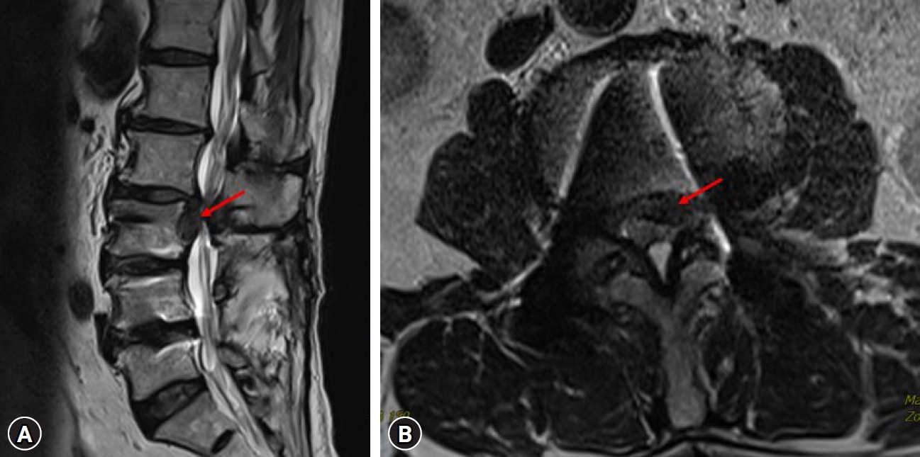
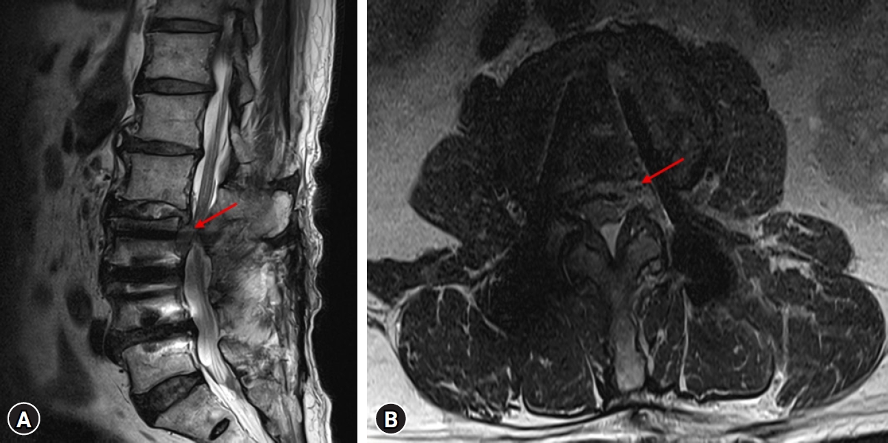
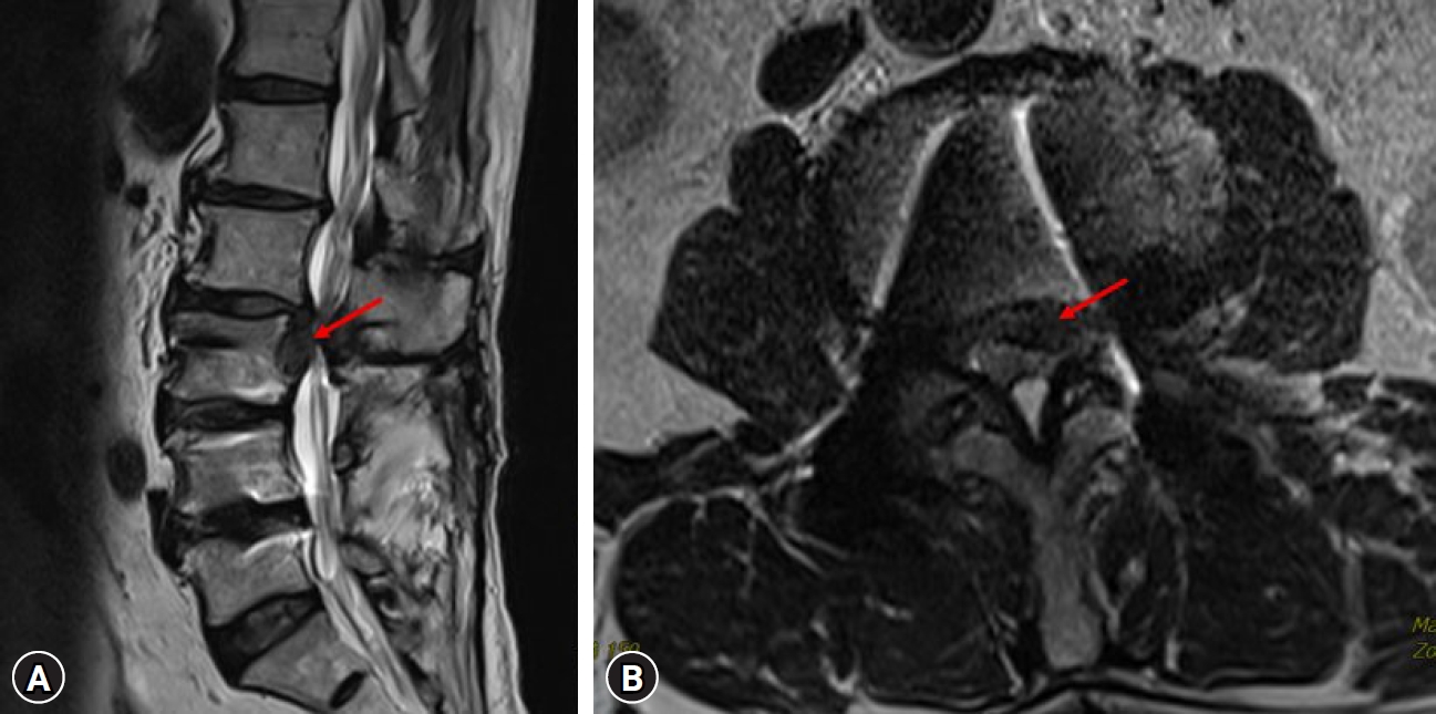
A 76-year-old male presented with progressive left lower extremity weakness and radicular pain. Imaging revealed a left-sided ruptured disc with inferior migration at the L2–3 level, causing compression of the left L3 nerve root (Fig. 1A, 1B). The patient underwent transforaminal endoscopic lumbar discectomy (TELD) at L2–3. Postoperatively, motor weakness improved; however, radicular pain along the L3 dermatome persisted despite adequate decompression and the absence of a definite residual compressive lesion on follow-up imaging (Fig. 2A, 2B).
A selective nerve root block (SNRB) targeting the left L3 nerve root resulted in temporary symptom relief. Based on this response, PRF followed by low-temperature thermal radiofrequency was performed. After the procedure, the patient reported significant pain reduction (visual analog scale [VAS] score decreased from 7 to 2), with mild transient numbness that resolved spontaneously. Symptom improvement was maintained for over 12 months without recurrence or complications.
2. Case 2
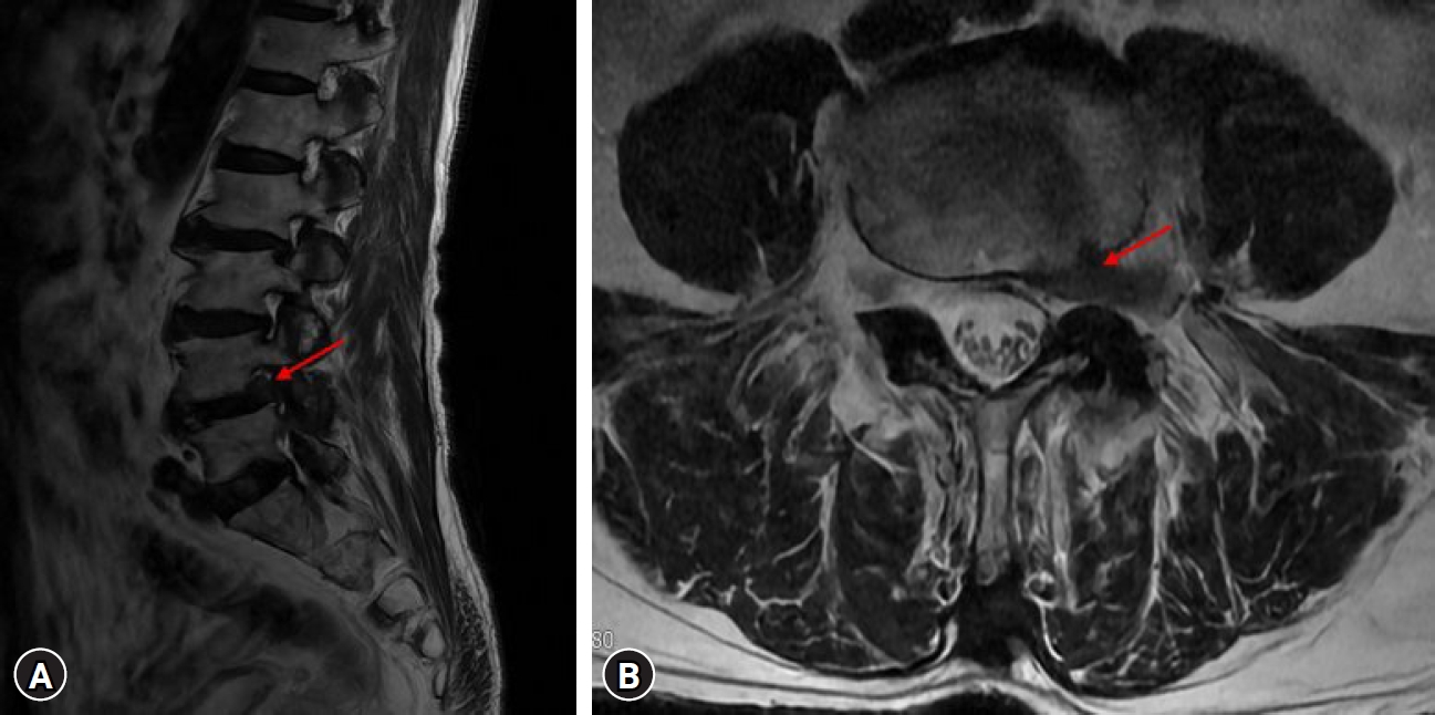
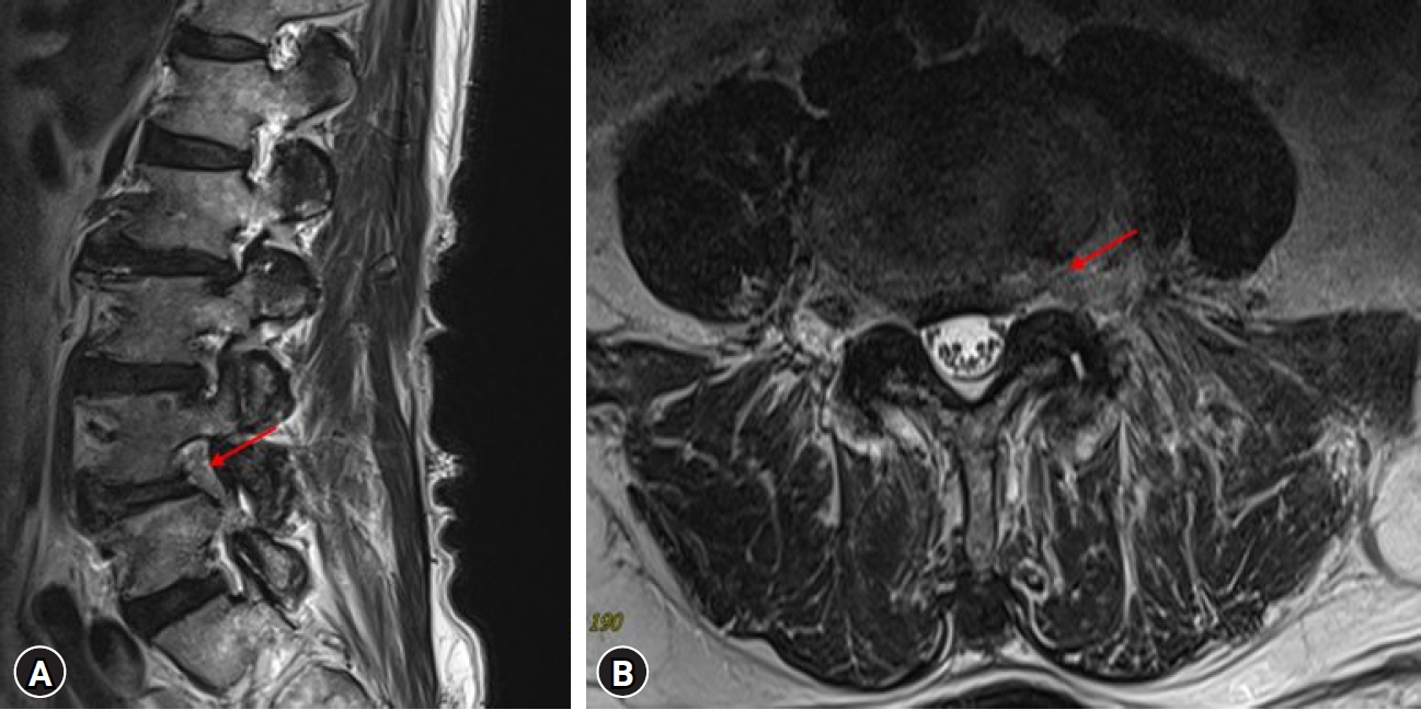
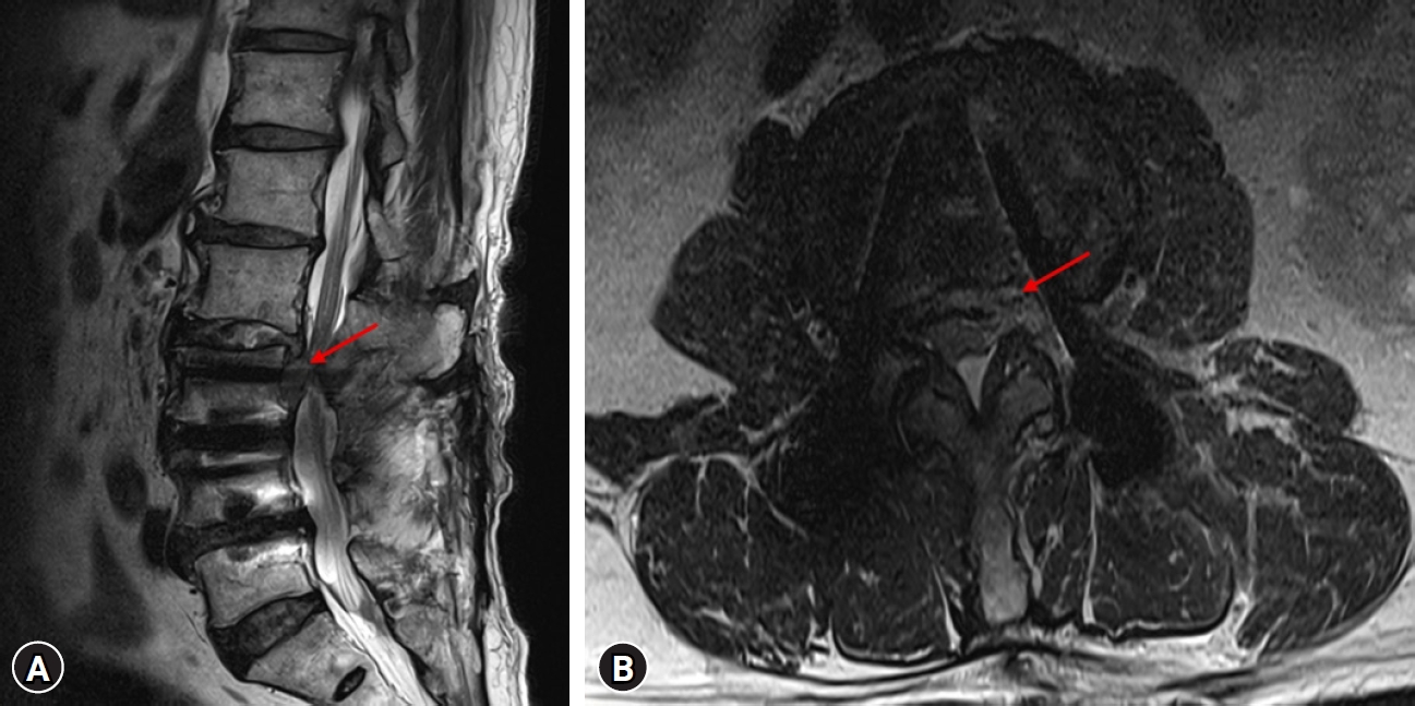
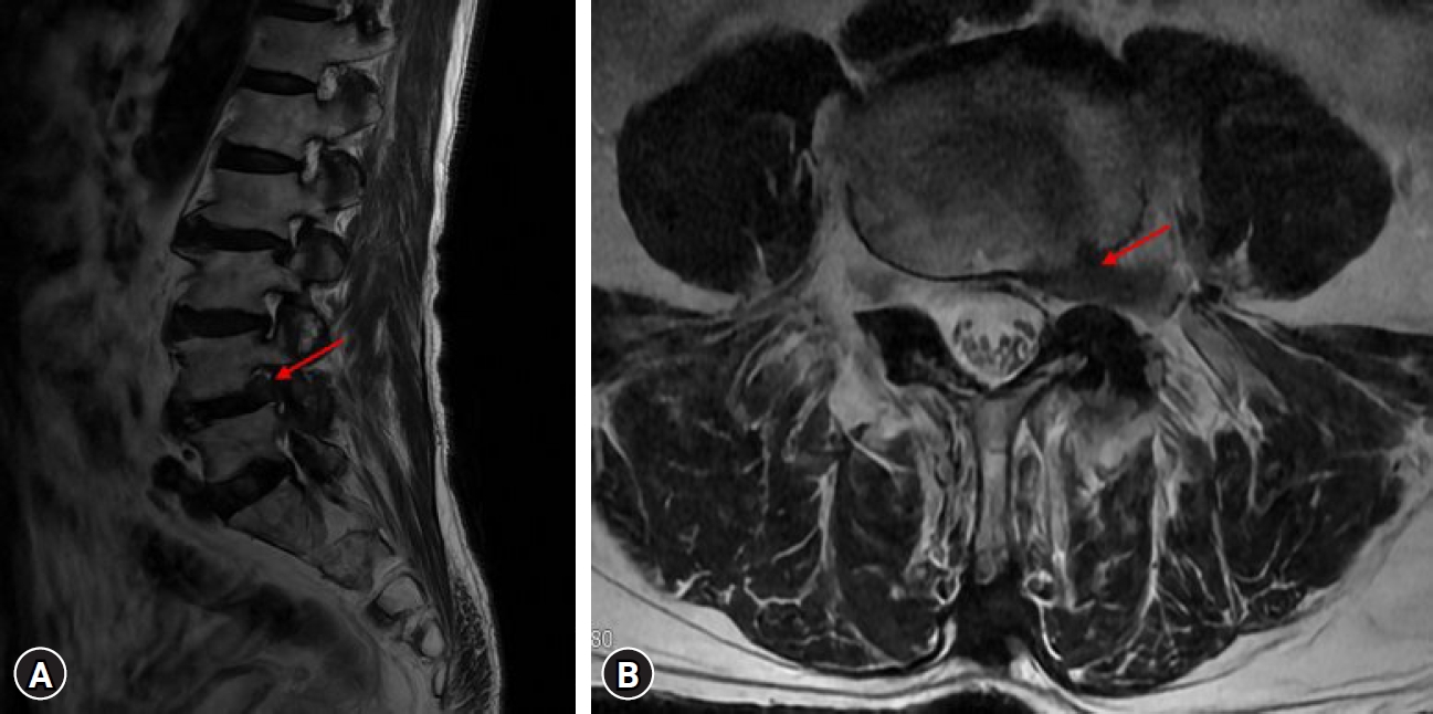
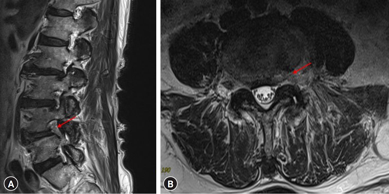
A 79-year-old male presented with left lower extremity radicular pain and motor weakness. Imaging revealed a ruptured disc at the L4–5 level causing compression of the left L4 nerve root (Fig. 3A, 3B). The patient underwent TELD at L4–5. Postoperatively, motor weakness improved; however, radicular pain along the L4 dermatome persisted despite adequate decompression and the absence of a definite residual compressive lesion on follow-up imaging (Fig. 4A, 4B).
A left L4 SNRB provided temporary symptom relief, but the pain recurred. After 3 months of conservative management, combined PRF and low-temperature thermal radiofrequency was performed. The patient reported significant pain reduction (VAS score decreased from 6 to 3), with transient numbness along the L4 distribution. At 2-year follow-up, symptom improvement was maintained without recurrence or complications.
3. Intervention technique
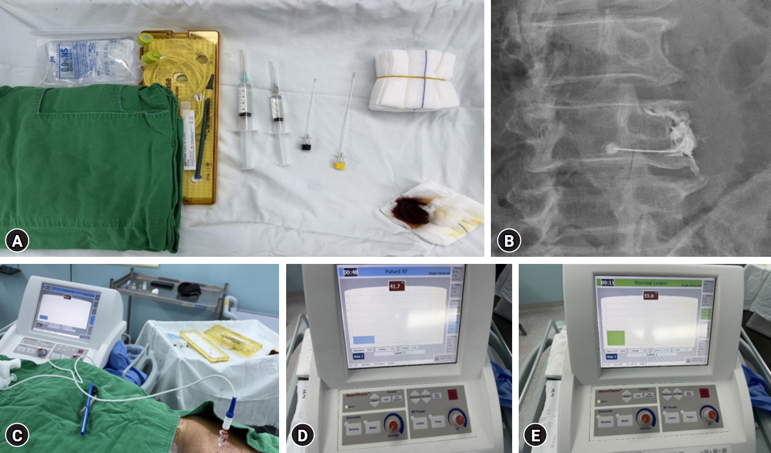
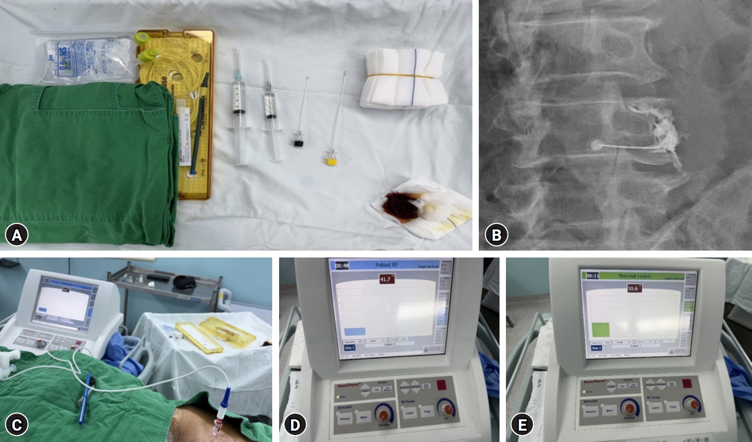
All instruments used for the procedure, including the radiofrequency (RF) needle and electrode, are shown in Fig. 5A. After sterile preparation and local anesthesia, an RF needle (22-gauge, 10 cm, straight, 5 mm active tip) was advanced to the target nerve root. Contrast injection was used to confirm accurate positioning of the needle tip (Fig. 5B). An RF electrode (10 cm reusable stainless-steel electrode) was then inserted through the needle and connected to the RF generator (IONIC RF Generator, Abbott Medical, St. Paul, MN, USA) (Fig. 5C). Sensory stimulation (monopolar, maximum voltage 3 V, frequency 50 Hz, pulse width 1 ms) was performed to confirm correct targeting of the symptomatic nerve root. PRF was applied first at a maximum temperature of 42°C for 120 seconds (monopolar, maximum voltage 45 V, frequency 2 Hz, pulse width 20 ms) (Fig. 5D). Subsequently, the system was switched to lesion mode, and low-temperature thermal radiofrequency was applied at a target temperature of 55°C for 60 seconds (monopolar) (Fig. 5E). After completion of the procedure, a total of 10 mL of mixed solution (2 mL of 2% lidocaine, 1 mL of dexamethasone, and 7 mL of normal saline) was injected around the target nerve root. Written informed consent for publication was obtained from the patients.
Discussion
Postoperative radicular pain is a complex condition that cannot be explained solely by mechanical compression. In the present cases, radicular symptoms were observed despite adequate decompression, suggesting the involvement of additional non-compressive mechanisms. Previous studies have shown that inflammatory mediators around the intervertebral disc can induce radiculitis, increasing nerve root sensitivity.1,2) Repeated or sustained neural irritation may also lead to hyperexcitability of the dorsal root ganglion (DRG), resulting in spontaneous neuronal firing and amplification of pain signals.8,9) In addition, neuropathic pain arising from abnormal neural signaling pathways plays a significant role in radicular symptoms.3) Therefore, the combined approach in this study was primarily aimed at modulating DRG sensitization and neuropathic pain components.
PRF is a neuromodulation technique that exerts its effect through electric fields rather than thermal nerve destruction, thereby minimizing structural damage while modulating pain transmission.6,8) It is believed to interfere with nociceptive signal transmission from peripheral nerves to the central nervous system.10) Although some studies have reported limited high-quality evidence supporting PRF, the reported benefits have often been limited to short-term efficacy.11-15) Other studies have shown clinically meaningful pain relief following PRF applied to the DRG in patients with radicular pain.4,8,16,17) A recent systematic review suggested that PRF may provide superior pain relief compared with lumbar epidural block at three months post-procedure, although the overall level of evidence remains limited.11)
According to experimental studies analyzing the phenomena occurring at the RF electrode tip, transient temperature elevations may occur during PRF application; however, these thermal effects are generally insufficient to achieve the level of ablation typically intended in conventional radiofrequency treatment.6,7,18) In contrast, conventional RF is usually applied at higher temperatures (80°C–90°C), inducing neural suppression through protein denaturation, but it carries a relatively higher risk of irreversible neural injury.6,7,18) In this context, we considered the addition of a controllable low-temperature thermal component to complement the neuromodulatory effects of PRF. At a relatively low target temperature of 55°C, this approach can be interpreted as a form of thermal modulation rather than conventional lesioning and may enhance clinical efficacy while maintaining procedural safety. Therefore, the combined application of PRF and low-temperature thermal RF may represent a rational and clinically applicable treatment strategy, integrating neuromodulatory effects with a controlled thermal component to overcome the limitations of each modality when used alone.
To the best of our knowledge, there are no previous reports describing the combined use of PRF and low-temperature thermal radiofrequency in this context, which represents a notable aspect of this study. Various treatment options have been proposed for postoperative radicular pain, including repeated nerve block, revision surgery, spinal cord stimulation (SCS), and DRG stimulation. Repeated nerve block may provide temporary symptom relief; however, recurrence of pain is common and long-term efficacy is often limited. Revision surgery may not be appropriate in the absence of a definite compressive lesion, while neuromodulation techniques such as SCS and DRG stimulation are relatively invasive and costly despite their demonstrated clinical efficacy in refractory neuropathic pain conditions.9,19) In this context, combined PRF and low-temperature thermal radiofrequency may represent a feasible minimally invasive alternative in carefully selected patients. This approach may be particularly applicable in patients with radicular pain who demonstrate a positive response to SNRB despite the absence of clear surgical indications. However, this report is limited by the small number of cases and the absence of a control group. Therefore, further studies are required to better define the appropriate indications for this procedure and to validate its safety and clinical effectiveness.
Combined PRF and low-temperature thermal radiofrequency may represent a feasible minimally invasive treatment option for postoperative radicular pain. Further studies are needed to clarify its effectiveness and indications.
The authors have no conflicts of interest to declare.
Funding
None.
Acknowledgments
None.
Fig. 1.
Preoperative magnetic resonance imaging sagittal view (A) and axial view (B) showing a left-sided disc herniation with inferior migration at the L2–3 level (arrows).
Fig. 2.
Postoperative magnetic resonance imaging sagittal view (A) and axial view (B) showing adequate decompression at the L2–3 level after endoscopic discectomy (arrows).
Fig. 3.
Preoperative magnetic resonance imaging sagittal view (A) and axial view (B) showing a left-sided herniation at the L4–5 level (arrows).
Fig. 4.
Postoperative magnetic resonance imaging sagittal view (A) and axial view (B) showing adequate decompression at the L4–5 level after endoscopic discectomy (arrows).
Fig. 5.
Procedure of pulsed radiofrequency (PRF) and low-temperature thermal radiofrequency performed in case 2. (A) Instruments used for the procedure, including the radiofrequency (RF) electrode, RF needle, and a 10-mL syringe containing a mixture of lidocaine and steroid. (B) Fluoroscopic image showing selective nerve root targeting and contrast confirmation. (C) Intra-procedural image showing the RF electrode connected to the RF generator (IONIC RF Generator, Abbott Medical) during treatment. (D) Generator display during PRF (42°C, 120 seconds). (E) Generator display during low-temperature thermal radiofrequency (55°C, 60 seconds).
References
1. Olmarker K, Rydevik B. Pathophysiology of sciatica. Orthop Clin North Am 1991;22:223-34.
2. McCarron RF, Wimpee MW, Hudkins PG, Laros GS. The inflammatory effect of nucleus pulposus: a possible element in the pathogenesis of low-back pain. Spine (Phila Pa 1976) 1987;12:760-4.
5. Sam J, Catapano M, Sahni S, Ma F, Abd-Elsayed A, Visnjevac O. Pulsed radiofrequency in interventional pain management: cellular and molecular mechanisms of action - an update and review. Pain Physician 2021;24:525-32.
9. Deer TR, Hunter CW, Mehta P, et al. A systematic literature review of dorsal root ganglion neurostimulation for the treatment of pain. Pain Med 2020;21:1581-9.
10. Abd-Elsayed A, Hughes M, Narel E, Loebertman MD. The efficacy of radiofrequency ablation for pain management in patients with pre-existing hardware at the site of ablation. Pain Ther 2020;9:709-16.
11. Park S, Park JH, Jang JN, et al. Pulsed radiofrequency of lumbar dorsal root ganglion for lumbar radicular pain: a systematic review and meta-analysis. Pain Pract 2024;24:772-85.
12. Shanthanna H, Chan P, McChesney J, Thabane L, Paul J. Pulsed radiofrequency treatment of the lumbar dorsal root ganglion in patients with chronic lumbar radicular pain: a randomized, placebo-controlled pilot study. J Pain Res 2014;7:47-55.
13. Vuka I, Dosenovic S, Marcius T, et al. Efficacy and safety of pulsed radiofrequency as a method of dorsal root ganglia stimulation for treatment of non-neuropathic pain: a systematic review. BMC Anesthesiol 2020;20:105.
17. Van Zundert J, Patijn J, Kessels A, Lame I, van Suijlekom H, van Kleef M. Pulsed radiofrequency adjacent to the cervical dorsal root ganglion in chronic cervical radicular pain: a double blind sham controlled randomized clinical trial. Pain 2007;127:173-82.
18. Perret DM, Kim DS, Li KW, et al. Application of pulsed radiofrequency currents to rat dorsal root ganglia modulates nerve injury-induced tactile allodynia. Anesth Analg 2011;113:610-6.
19. North RB, Kidd DH, Farrokhi F, Piantadosi SA. Spinal cord stimulation versus repeated lumbosacral spine surgery for chronic pain: a randomized, controlled trial. Neurosurgery 2005;56:98-106.
Clinical Application of Combined Pulsed Radiofrequency and Low-Temperature Thermal Radiofrequency for Postoperative Radicular Pain: A Case Report
Fig. 1. Preoperative magnetic resonance imaging sagittal view (A) and axial view (B) showing a left-sided disc herniation with inferior migration at the L2–3 level (arrows).
Fig. 2. Postoperative magnetic resonance imaging sagittal view (A) and axial view (B) showing adequate decompression at the L2–3 level after endoscopic discectomy (arrows).
Fig. 3. Preoperative magnetic resonance imaging sagittal view (A) and axial view (B) showing a left-sided herniation at the L4–5 level (arrows).
Fig. 4. Postoperative magnetic resonance imaging sagittal view (A) and axial view (B) showing adequate decompression at the L4–5 level after endoscopic discectomy (arrows).
Fig. 5. Procedure of pulsed radiofrequency (PRF) and low-temperature thermal radiofrequency performed in case 2. (A) Instruments used for the procedure, including the radiofrequency (RF) electrode, RF needle, and a 10-mL syringe containing a mixture of lidocaine and steroid. (B) Fluoroscopic image showing selective nerve root targeting and contrast confirmation. (C) Intra-procedural image showing the RF electrode connected to the RF generator (IONIC RF Generator, Abbott Medical) during treatment. (D) Generator display during PRF (42°C, 120 seconds). (E) Generator display during low-temperature thermal radiofrequency (55°C, 60 seconds).
Fig. 1.
Fig. 2.
Fig. 3.
Fig. 4.
Fig. 5.
Clinical Application of Combined Pulsed Radiofrequency and Low-Temperature Thermal Radiofrequency for Postoperative Radicular Pain: A Case Report


 , Jong Hun Seo, M.D., Ph.D.