Department of Neurosurgery, Inha University College of Medicine, Incheon, Korea
Corresponding author: Seung-Hwan Yoon, M.D., Ph.D. Department of Neurosurgery, Inha University College of Medicine, 27 Inhang-ro, Jung-gu, Incheon, Korea TEL: +82-32-890-2370, FAX: +82-32-890-2374 E-mail: nsyoon@gmail.com
• Received: May 29, 2026 • Revised: June 2, 2026 • Accepted: June 4, 2026
This is an open access article distributed under the terms of the Creative Commons Attribution Non-Commercial License (https://creativecommons.org/licenses/by-nc/4.0/) which permits unrestricted non-commercial use, distribution, and reproduction in any medium, provided the original work is properly cited.
This retrospective study investigated the distinct clinical and radiographic drivers of early- versus late-onset proximal junctional kyphosis (PJK) following multilevel thoracolumbar (TL) fusion.
Methods
After applying the exclusion criteria (spinal infection, neuromuscular disease, age <50 years), the analysis included 136 patients who underwent ≥4-level TL fusion and were followed up for a minimum of 2 years. PJK was classified as early (≤6 months) or late (>6 months) onset. Patient-related factors, surgical variables, sagittal spinopelvic parameters, and preoperative magnetic resonance imaging findings were analyzed using multivariate logistic regression to identify independent predictors of early PJK.
Results
Among 24 patients (17.6%) who developed PJK, the early and late-onset groups included 13 and 11 patients, respectively. The early PJK group exhibited significantly greater preoperative and postoperative TL angles compared with the late group (preoperative: 23.03±13.83° vs. 9.67±9.67°, p=0.024; postoperative: 19.6±6.95° vs. 6.95±6.35°, p<0.001). The Pfirrmann grade of the L1–2 intervertebral disc was significantly higher in the early PJK group (3.92±0.95 vs. 2.81±0.60, p=0.006). No surgical variables differed significantly between the groups. Multivariate analysis confirmed greater postoperative TL angle and more advanced L1–2 disc degeneration as independent predictors of early PJK.
Conclusion
Early-onset PJK following multilevel TL fusion is primarily driven by regional biomechanical vulnerabilities, specifically residual postoperative TL kyphosis and advanced adjacent L1–2 disc degeneration, rather than by surgical variables. Meticulous evaluation of regional TL alignment and adjacent disc health during surgical planning is critical for risk stratification and prevention of early junctional failure.
Multilevel thoracolumbar (TL) spinal fusion is increasingly performed for the management of advanced degenerative spinal disease, spinal deformity, and vertebral fractures, particularly in patients >60 years.1)
The complications of multilevel spinal fusion include adjacent segment disease, proximal junctional kyphosis (PJK), and proximal junctional failure (PJF). Among these, PJK is a postoperative radiographic phenomenon occurring after surgery for spinal deformities in both adult and pediatric populations and is generally considered a form of adjacent segment pathology.2) PJK is commonly defined as a proximal junctional angle (PJA) of >10° and at least 10° greater than the preoperative measurement between the upper instrumented vertebra (UIV) and the two supra-adjacent vertebrae.3,4) PJF is a more severe condition within the spectrum of PJK and is characterized by structural failure, including vertebral body fracture or posterior ligamentous disruption, which may lead to pain or neurological deficits.5)
Although many patients with radiographic PJK remain asymptomatic, some patients with progressive deformities or neurological deterioration may require revision surgery. The risk factors for PJK include advanced age, preoperative sagittal imbalance, excessive sagittal correction, combined anteroposterior approaches, sacral fusion, low bone mineral density (BMD), and a high body mass index (BMI).2,4,6,7)
PJK is frequently identified during the early postoperative period, particularly within the first 6 months following multilevel spinal fusion, a threshold that has been widely employed to capture the early acute biomechanical phase of junctional failure and often presents a challenging clinical course. Despite this observation, most previous studies have analyzed PJK as a single entity without distinguishing between early- and late-onset patterns. Consequently, factors specifically associated with early-onset PJK remain insufficiently characterized.
The TL junction is a biomechanically vulnerable transition zone, in which altered sagittal alignment and adjacent disc degeneration may increase mechanical stress at the proximal junction following long-segment fusion.
Therefore, the present study compared early- and late-onset PJK following multilevel TL fusion and identified factors specifically associated with early PJK, with particular attention paid to sagittal alignment parameters and preoperative magnetic resonance imaging (MRI) findings.
Methods
1. Study design and patient population
We retrospectively reviewed the records of 141 patients who underwent TL fusion involving ≥4 vertebrae between April 2011 and August 2020. All surgeries were performed by a single spinal surgeon at a single institution. The minimum postoperative follow-up period was 2 years, and the mean follow-up duration was 3.6 years.
The inclusion criteria were as follows: (1) age >50 years at the time of surgery; (2) diagnosis of degenerative spinal disease, including spinal stenosis, spondylolisthesis, or osteoporotic vertebral compression fracture; and (3) multilevel TL fusion involving ≥4 levels. Patients were excluded if they had a spinal infection (n=2) or neuromuscular disease, including Parkinsonism (n=1), or were <50 years of age (n=2). After applying the exclusion criteria, 136 patients were eligible for inclusion in the final analysis.
The baseline patient characteristics included age, sex, height, weight, BMI, smoking status, and BMD. BMD was measured at the lumbar spine and femur using dual-energy X-ray absorptiometry (Prodigy, GE Healthcare, Madison, WI, USA).
Among these patients, 24 (17.6%) were diagnosed with PJK during follow-up. Patients who developed PJK were further classified according to the timing of the onset into early PJK (occurring ≤6 months postoperatively, n=13) and late PJK (occurring >6 months postoperatively, n=11), based on the 6-month cutoff commonly used in the literature to distinguish acute biomechanical failure from delayed degenerative junctional failure. Postoperative follow-up evaluations were routinely performed at 1 week; 1, 3, and 6 months after surgery, and at 6-month intervals thereafter.
2. Radiologic evaluation
1) Lumbar MRI
Preoperative lumbar MRI was performed using a 1.5-T system (Signa HDxt, GE Medical Systems, Milwaukee, WI, USA). Degenerative changes were evaluated using sagittal T1- and T2-weighted images. The presence of disc protrusion or extrusion, moderate-to-severe central canal stenosis, compression fracture (vertebral height loss was classified as mild, 20–25%; moderate, 25–40%; or severe, >40%), and spondylolisthesis (Meyerding grades I–II) was assessed.
Intervertebral disc degeneration at each lumbar level was graded using the Pfirrmann classification system.8) To evaluate fatty infiltration of the paraspinal muscles, muscle quality was graded according to the Goutallier classification as follows: grade 0, normal muscle; grade 1, fatty streaks within the muscle; grade 2, less fat than muscle; grade 3, equal amounts of fat and muscle; and grade 4, more fat than muscle.9)
2) Sagittal spinopelvic parameters
Sagittal spinopelvic parameters were measured using standing whole-spine lateral radiographs obtained preoperatively and 1 week postoperatively. The evaluated parameters included sagittal vertical axis, thoracic kyphosis, lumbar lordosis (LL), TL kyphosis, pelvic incidence (PI), pelvic tilt, sacral slope, and PI-LL mismatch.10)
Thoracic kyphosis was defined as the Cobb angle between the upper endplate of T5 and the lower endplate of T12. Lumbar lordosis was measured between the lower end plate of T12 and the upper end plate of S1. TL kyphosis was defined as the Cobb angle between the upper endplate of T10 and the lower endplate of L2. Pelvic parameters were measured using standard definitions. Preoperative and postoperative values, as well as changes in each parameter, were compared between the early and late PJK groups.
3) PJK analysis
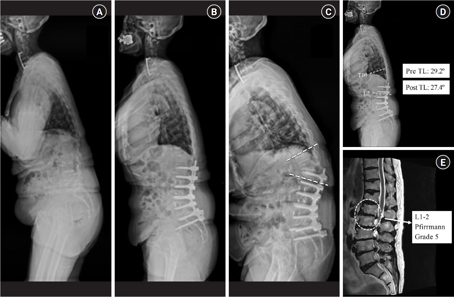
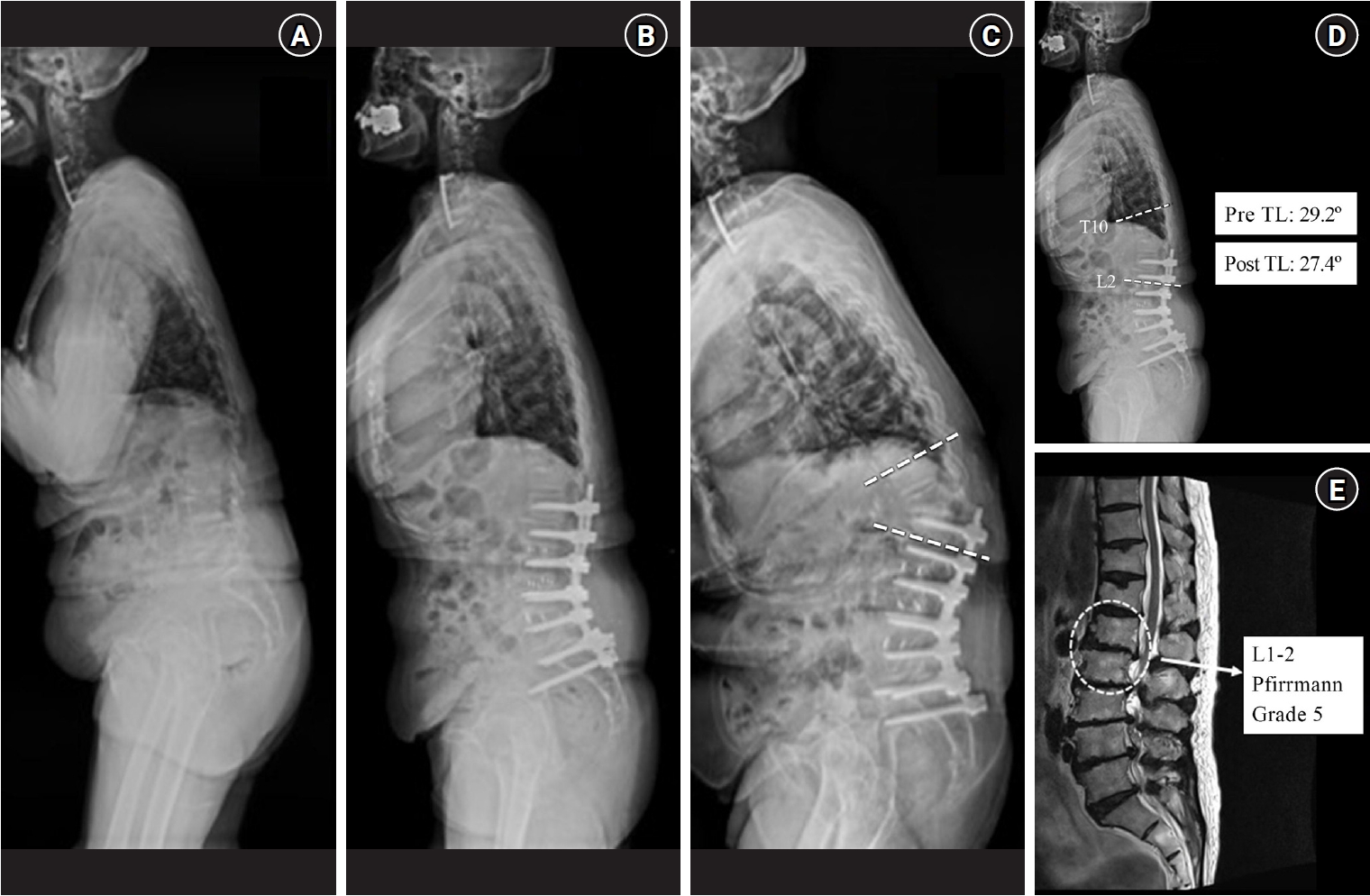
On each follow-up radiograph, the PJA was measured between the caudal endplate of the UIV and the cranial endplate of the second supra-adjacent vertebra. PJK was defined as an increase in the PJA of at least 10° compared with the preoperative measurement.11) The presence of PJK and the time to its development were recorded for all patients (Fig. 1A–C).
3. Surgical factors
The surgical variables analyzed in this study included a history of previous spinal fusion, surgical approach (posterior-only or combined anterior-posterior), use of iliac screw fixation, level of the UIV above T9, level of the lowest instrumented vertebra (LIV at S1), number of fused levels, osteotomy performance (pedicle subtraction or Smith–Petersen osteotomy), UIV/LIV Cobb angle, and cement augmentation at the proximal junctional segments, including cement-augmented pedicle screws or percutaneous vertebroplasty.
4. Statistical analysis
Descriptive data are presented as means±standard deviations for continuous variables and as frequencies and percentages for categorical variables. The normality of continuous variables was assessed using the Shapiro-Wilk test. The early and late PJK groups were compared using the chi-square test, Student’s t-test, or Mann-Whitney U test, as appropriate. Variables significant in the univariate analysis were entered into the multivariate logistic regression model to identify independent factors associated with early PJK. Statistical significance was set at p<0.05. All statistical analyses were performed using SPSS version 26.0 (IBM Corp., Armonk, NY, USA).
5. Ethics statement
This retrospective cohort study was approved by the Institutional Review Board (IRB No. 2022-08-012-000). This type of study does not require informed consent.
Results
1. Baseline characteristics
The baseline characteristics of the patients with early and late PJK are summarized in Table 1. The mean age of patients did not differ significantly between the early and late PJK groups (68.92±6.72 years vs. 69.0±6.37 years, p=0.977). Female patients were predominant in both groups, with no significant difference in sex distribution between the groups (p=0.202).
Height, weight, BMI, smoking status, and BMD measured at the lumbar spine or femur did not differ significantly between the groups.
2. PJK characteristics and timing
The modes of PJK according to onset group are summarized in Table 1. The mean time to PJK onset in all patients was 18 months after surgery. In the early PJK group, PJK developed at a mean of 3 months postoperatively, whereas the mean onset time in the late PJK group was 35 months postoperatively.
Regarding the mode of PJK failure, vertebral fracture at the UIV or adjacent supra-adjacent vertebra was the most prevalent pattern in both groups. Fracture-related PJK was more frequently observed in the early PJK group than in the late PJK group, although the difference was not statistically significant.
3. Risk factor comparison
Comparisons of the surgical variables between the early and late PJK groups are summarized in Table 1. No statistically significant differences were observed between the two groups with respect to prior spinal surgery, surgical approach, use of iliac screw fixation, level of the UIV or LIV, number of fused levels, osteotomy performance, UIV/LIV Cobb angle, or use of cement augmentation.
Overall, none of the analyzed surgical variables were significantly associated with the timing of PJK onset.
4. Sagittal alignment and spinopelvic parameters
The pre- and postoperative sagittal alignment and spinopelvic parameters are summarized in Table 2. Preoperatively, the TL angle was significantly greater in the early PJK group compared with the late PJK group (23.03±13.83° vs. 9.67±9.67°, p=0.024). No other preoperative sagittal parameters differed significantly between the groups.
Postoperatively, the TL angle remained significantly higher in the early PJK group than in the late PJK group (19.6±6.95° vs. 6.95±6.35°, p<0.001). Other postoperative sagittal and pelvic parameters did not differ significantly between the two groups.
Comparison of the changes in sagittal parameters from the preoperative to the postoperative period showed that although the change in sagittal vertical axis tended to be greater in the late PJK group (31.34±77.33 mm vs. 8.11±30.56 mm, p=0.329), this difference was not statistically significant. No significant intergroup differences were observed in other alignment parameters.
5. MRI findings
Comparisons of preoperative lumbar MRI findings between the early and late PJK groups are shown in Table 3. The prevalence of disc protrusion or extrusion, central canal stenosis, spondylolisthesis, and compression fractures did not differ significantly between the groups. Fatty infiltration of the paraspinal muscles was also comparable.
In contrast, the degree of intervertebral disc degeneration at the L1–2 level, as assessed using the Pfirrmann grading system, was significantly greater in the early PJK group than in the late PJK group (3.92±0.95 vs. 2.81±0.60, p=0.006). Pfirrmann grades did not differ significantly at other lumbar levels.
6. Multivariate logistic regression analysis
The results of the multivariate logistic regression analysis are presented in Table 4. A representative case illustrating these findings is shown in Fig. 1D and 1E. The model included variables demonstrating potential associations in univariate analysis.
In the final multivariate model, a greater postoperative TL angle and a higher Pfirrmann grade of the L1–2 intervertebral disc were independently associated with early PJK. No surgical or other radiographic variables were significant predictors in the multivariate analysis.
Discussion
PJK remains one of the most challenging complications of long-segment TL spinal fusion.11) The reported incidence of PJK ranges from 20% to 35% in adult spinal deformity surgeries, with a pooled incidence of approximately 30%.4) In the present study, PJK developed in 17.6% of patients after multilevel spinal fusion, within the lower range of previous reports. Among the patients who developed PJK, the temporal patterns and clinical characteristics differed between early- and late-onset PJK.
Early-onset PJK frequently develops within the first few months after surgery.7,12) Yagi et al.7) reported that approximately 76% of PJK cases occurred within 3 months. Consistent with this finding, early PJK in the present study occurred at a mean of 3 months postoperatively, whereas late PJK developed at a mean of 35 months. These temporal patterns support the clinical relevance of distinguishing early- from late-onset PJK, because early-onset PJK likely reflects acute biomechanical failure, whereas late-onset PJK may represent progressive degenerative changes.
The patient-related risk factors previously associated with PJK include advanced age, female sex, high BMI, and low BMD.2,4,6,13,14) Osteopenia and osteoporosis have been suggested to contribute to junctional failure by reducing vertebral load-bearing capacity, particularly in older patients.6,15-17) In the present study, the baseline demographic characteristics and BMD did not differ significantly between patients with early and late PJK. Although compression fractures were more frequently observed in the early PJK group, the difference was not statistically significant. These findings suggest that patient-related factors alone may not adequately explain the timing of PJK onset.
The surgical factors implicated in PJK development include surgical approach, UIV/LIV level selection, fusion length, osteotomy, rod stiffness, and cement augmentation.3,7,14,18-21) In particular, fusion constructs extending to the sacrum are associated with higher rates of junctional complications.22) However, in the present study, none of the analyzed surgical variables, including surgical approach, UIV and LIV levels, fusion length, osteotomy, iliac fixation, or cement augmentation, differed significantly between the early and late PJK groups. This finding suggests that surgical factors alone may not be the primary determinants of early PJK onset in this cohort.
In contrast, the results of sagittal alignment analysis demonstrated that TL kyphosis was significantly associated with early PJK. Both preoperative and postoperative TL angles were significantly greater in the early PJK group compared with the late PJK group, whereas other global sagittal and spinopelvic parameters did not differ significantly between the groups. Although the preoperative TL angle showed significance in univariate analysis, it did not remain significant in the multivariate model, suggesting that postoperative TL alignment, rather than baseline morphology alone, is more critical in the development of early PJK. Furthermore, the high collinearity between the pre- and postoperative TL angles likely contributed to the exclusion of the preoperative TL angle from the final multivariate model. Persistence of postoperative TL kyphosis likely increases mechanical stress at the proximal junction, thereby contributing to early structural failure. Moreover, the transition from a rigid thoracic spine to a more mobile lumbar segment can increase mechanical stress at the proximal junction after long-segment fusion, particularly in the presence of residual TL kyphosis or dynamic motion-related factors.23) In the present study, the early PJK group showed significantly more advanced degeneration of the L1–2 intervertebral disc, as assessed by the Pfirrmann classification. This degeneration remained the strongest independent factor associated with early PJK in multivariate analysis (Table 4). Given the small subgroup size, however, the wide confidence interval (CI) for this estimate indicates that the magnitude of this association should be interpreted with caution. The L1–2 disc is located adjacent to the TL junction and is subjected to increased mechanical demand following long-segment fusion. Preexisting disc degeneration at this level may reduce resistance to kyphotic and compressive forces, thereby predisposing patients to early junctional failure. These findings are consistent with those reported previously, suggesting that adjacent segment degeneration near the TL junction increases the risk of junctional complications. Our results suggest that the health of the L1–2 intervertebral disc should be a primary consideration when selecting the UIV. In clinical practice, if preoperative MRI reveals advanced degeneration (Pfirrmann grade ≥4) at the L1–2 level, extending the fusion more cranially to a healthier segment may be considered to avoid placing the proximal junction at a biomechanically compromised level. However, because this suggestion is based on retrospective observations and was not directly tested in the present study, it should be regarded as hypothesis-generating and requires confirmation in prospective studies. Collectively, increased postoperative TL angle and advanced L1–2 disc degeneration may predispose patients to proximal vertebral instability, thereby facilitating early-onset PJK.3)
Several strategies have been proposed to reduce the risk of PJK and PJF, including cement augmentation, use of hooks at the UIV, and the use of transitional rods.18,24) However, these surgical techniques were not associated with differences in the timing of PJK onset in the present study. Instead, the findings emphasized the importance of careful consideration of TL alignment and adjacent disc condition during preoperative planning and intraoperative correction. Thus, patients with excessive TL kyphosis or advanced L1–2 disc degeneration may represent a subgroup at higher risk for early PJK.
The biomechanical role of L1–2 disc degeneration likely differs depending on whether L1–2 is included in the fusion construct. Disc degeneration at this level is unlikely to directly drive early PJK. When unfused, preexisting degeneration may compromise load-sharing and resistance to kyphotic forces, particularly with residual postoperative TL kyphosis. The association between L1–2 disc degeneration and early PJK in this study likely reflects adjacent-segment vulnerability at the TL junction, rather than a single isolated mechanism.
This study had several limitations. First, the retrospective design and small sample size may have limited the generalizability of the results. In particular, the restricted number of patients in each subgroup (early PJK, n=13; late PJK, n=11) may have resulted in an underpowered multivariate model, and the wide CI observed for the L1–2 Pfirrmann grade (odds ratio, 22.6; 95% CI, 1.667 to 306.3) should be interpreted with caution, as it may reflect statistical instability rather than a true effect magnitude. Accordingly, the odds ratio for the L1–2 Pfirrmann grade should be regarded as imprecise and interpreted with particular caution, rather than as a precise estimate of effect size. Second, detailed bone quality assessments beyond BMD were unavailable. Third, the follow-up period may not have fully captured late-onset junctional complications.
Early-onset PJK after multilevel TL fusion is uniquely associated with greater postoperative TL kyphosis and more advanced degeneration of the adjacent L1–2 intervertebral discs. Conversely, surgical factors and global sagittal alignment parameters were not significantly associated with PJK onset. These findings indicate that early PJK is more closely associated with regional sagittal alignment at the biomechanically vulnerable TL junction and the preexisting structural integrity of the adjacent intervertebral disc, rather than to surgical techniques alone. Therefore, meticulous correction of regional TL alignment and thorough preoperative evaluation of the adjacent disc condition are important for identifying and managing patients at a high risk of early PJK.
NOTES
Author contributions
Conceptualization: SHY. Methodology: DSR, SHY. Data curation: WSK. Formal analysis: WSK, DSR. Investigation: WSK, DSR. Supervision: SHY. Writing – original draft: WSK. Writing – review & editing: DSR, SHY. All authors read and approved the final manuscript.
Conflict of interest
The authors have no conflicts of interest to declare.
Funding
None.
Acknowledgments
None.
Fig. 1.
Representative case of early proximal junctional kyphosis (PJK) in a 72-year-old woman with a stooping gait. (A) Preoperative standing lateral radiograph demonstrating degenerative flatback deformity. (B) Immediate postoperative radiograph following multi-level thoracolumbar fusion. (C) Follow-up radiograph obtained 3 months postoperatively demonstrating early PJK, with dashed lines indicating the increased proximal junctional angle. (D) Postoperative standing lateral radiograph with annotated T10–L2 Cobb angle measurement, demonstrating persistently elevated thoracolumbar kyphosis (preoperative TL angle: 29.2°; postoperative thoracolumbar [TL] angle: 27.4°). (E) Preoperative sagittal magnetic resonance imaging demonstrating advanced degeneration of the L1–2 intervertebral disc (Pfirrmann grade V, dashed circle).
Table 1.
Comparison of baseline characteristics, surgical variables, and mode of proximal junctional kyphosis between the early and late PJK groups
Early PJK (n=13)
Late PJK (n=11)
p-value
Age (years)
68.92±6.72
69±6.37
0.977
Male:Female
4:9
1:10
0.202
Height (cm)
150.57±8.39
151.48±5.76
0.766
Weight (kg)
55.53±6.94
54.43±9.33
0.977
BMI
24.53±2.61
23.61±3.42
0.467
Current smoking
3 (23.1)
2 (18.2)
0.773
Lumbar BMD
–1.37±0.94
–1.59±1.85
0.718
Femoral BMD
–2.05±0.95
–1.89±1.24
0.772
PJK mode
Fracture of UIV or UIV+1
12 (92.3)
8 (72.7)
0.300
Instrumented failure
1 (7.7)
3 (27.3)
Surgical variables
Previous surgery
9 (69.2)
10 (90.9)
0.202
Posterior only approach
12 (92.3)
10 (90.9)
0.904
Iliac screw fixation
3 (23.1)
3 (27.3)
0.817
UIV (above T9)
2 (15.4)
3 (27.3)
0.496
UIV (T10–L2)
11 (84.6)
8 (72.7)
LIV (sacrum 1)
7 (53.8)
8 (72.7)
0.351
UIV/LIV cobb angle (°)
28.08±14.14
25.78±14.38
0.697
Posterior fusion levels
6.46±1.76
6.54±2.42
0.923
PSO
2 (15.4)
2 (18.2)
0.858
SPO
1 (7.7)
1 (9.1)
0.904
Cement screw or PVP
4 (30.8)
5 (45.5)
0.459
Values are presented as mean±standard deviation or number (%). PJK: proximal junctional kyphosis, BMI: body mass index, BMD: bone mineral density, UIV: upper instrumented vertebra, LIV: lowest instrumented vertebra, PSO: pedicle subtraction osteotomy, SPO: Smith-Petersen osteotomy, PVP: percutaneous vertebroplasty.
Table 2.
Comparison of sagittal alignment and pelvic parameters between the early PJK and late PJK groups
1. Rajaee SS, Bae HW, Kanim LE, Delamarter RB. Spinal fusion in the United States: analysis of trends from 1998 to 2008. Spine (Phila Pa 1976) 2012;37:67-76.
2. Kim DK, Kim JY, Kim DY, Rhim SC, Yoon SH. Risk factors of proximal junctional kyphosis after multilevel fusion surgery: more than 2 years follow-up data. J Korean Neurosurg Soc 2017;60:174-80.
3. Glattes RC, Bridwell KH, Lenke LG, Kim YJ, Rinella A, Edwards C 2nd. Proximal junctional kyphosis in adult spinal deformity following long instrumented posterior spinal fusion: incidence, outcomes, and risk factor analysis. Spine (Phila Pa 1976) 2005;30:1643-9.
5. Lau D, Clark AJ, Scheer JK, et al. Proximal junctional kyphosis and failure after spinal deformity surgery: a systematic review of the literature as a background to classification development. Spine (Phila Pa 1976) 2014;39:2093-102.
7. Yagi M, King AB, Boachie-Adjei O. Incidence, risk factors, and natural course of proximal junctional kyphosis: surgical outcomes review of adult idiopathic scoliosis. Minimum 5 years of follow-up. Spine (Phila Pa 1976) 2012;37:1479-89.
8. Pfirrmann CW, Metzdorf A, Zanetti M, Hodler J, Boos N. Magnetic resonance classification of lumbar intervertebral disc degeneration. Spine (Phila Pa 1976) 2001;26:1873-8.
11. Park SJ, Lee CS, Chung SS, Lee JY, Kang SS, Park SH. Different risk factors of proximal junctional kyphosis and proximal junctional failure following long instrumented fusion to the sacrum for adult spinal deformity: survivorship analysis of 160 patients. Neurosurgery 2017;80:279-86.
12. Annis P, Lawrence BD, Spiker WR, et al. Predictive factors for acute proximal junctional failure after adult deformity surgery with upper instrumented vertebrae in the thoracolumbar spine. Evid Based Spine Care J 2014;5:160-2.
13. Bridwell KH, Lenke LG, Cho SK, et al. Proximal junctional kyphosis in primary adult deformity surgery: evaluation of 20 degrees as a critical angle. Neurosurgery 2013;72:899-906.
14. Kim HJ, Lenke LG, Shaffrey CI, Van Alstyne EM, Skelly AC. Proximal junctional kyphosis as a distinct form of adjacent segment pathology after spinal deformity surgery: a systematic review. Spine (Phila Pa 1976) 2012;37(22 Suppl):S144-64.
15. Echt M, Ranson W, Steinberger J, Yassari R, Cho SK. A systematic review of treatment strategies for the prevention of junctional complications after long-segment fusions in the osteoporotic spine. Global Spine J 2021;11:792-801.
16. Hostin R, McCarthy I, O'Brien M, et al. Incidence, mode, and location of acute proximal junctional failures after surgical treatment of adult spinal deformity. Spine (Phila Pa 1976) 2013;38:1008-15.
17. Watanabe K, Lenke LG, Bridwell KH, Kim YJ, Koester L, Hensley M. Proximal junctional vertebral fracture in adults after spinal deformity surgery using pedicle screw constructs: analysis of morphological features. Spine (Phila Pa 1976) 2010;35:138-45.
18. Ghobrial GM, Eichberg DG, Kolcun JP, et al. Prophylactic vertebral cement augmentation at the uppermost instrumented vertebra and rostral adjacent vertebra for the prevention of proximal junctional kyphosis and failure following long-segment fusion for adult spinal deformity. Spine J 2017;17:1499-505.
19. Mendoza-Lattes S, Ries Z, Gao Y, Weinstein SL. Proximal junctional kyphosis in adult reconstructive spine surgery results from incomplete restoration of the lumbar lordosis relative to the magnitude of the thoracic kyphosis. Iowa Orthop J 2011;31:199-206.
20. O'Leary PT, Bridwell KH, Lenke LG, et al. Risk factors and outcomes for catastrophic failures at the top of long pedicle screw constructs: a matched cohort analysis performed at a single center. Spine (Phila Pa 1976) 2009;34:2134-9.
21. Yagi M, Fujita N, Tsuji O, et al. Low bone mineral density is a significant risk factor for proximal junctional failure after surgical correction of adult spinal deformity: a propensity score-matched analysis. Spine (Phila Pa 1976) 2018;43:485-91.
22. Maruo K, Ha Y, Inoue S, et al. Predictive factors for proximal junctional kyphosis in long fusions to the sacrum in adult spinal deformity. Spine (Phila Pa 1976) 2013;38:E1469-76.
23. Ham DW, Han H, Kim HJ, Park SM, Chang BS, Yeom JS. Risk factors for acute proximal junctional kyphosis after adult spinal deformity surgery in preoperative motion analysis. Eur Spine J 2021;30:1215-25.
Beyond Surgical Factors: Postoperative Thoracolumbar Alignment and L1–2 Disc Degeneration as Independent Drivers of Early-Onset Proximal Junctional Kyphosis
Fig. 1. Representative case of early proximal junctional kyphosis (PJK) in a 72-year-old woman with a stooping gait. (A) Preoperative standing lateral radiograph demonstrating degenerative flatback deformity. (B) Immediate postoperative radiograph following multi-level thoracolumbar fusion. (C) Follow-up radiograph obtained 3 months postoperatively demonstrating early PJK, with dashed lines indicating the increased proximal junctional angle. (D) Postoperative standing lateral radiograph with annotated T10–L2 Cobb angle measurement, demonstrating persistently elevated thoracolumbar kyphosis (preoperative TL angle: 29.2°; postoperative thoracolumbar [TL] angle: 27.4°). (E) Preoperative sagittal magnetic resonance imaging demonstrating advanced degeneration of the L1–2 intervertebral disc (Pfirrmann grade V, dashed circle).
Fig. 1.
Beyond Surgical Factors: Postoperative Thoracolumbar Alignment and L1–2 Disc Degeneration as Independent Drivers of Early-Onset Proximal Junctional Kyphosis
Early PJK (n=13)
Late PJK (n=11)
p-value
Age (years)
68.92±6.72
69±6.37
0.977
Male:Female
4:9
1:10
0.202
Height (cm)
150.57±8.39
151.48±5.76
0.766
Weight (kg)
55.53±6.94
54.43±9.33
0.977
BMI
24.53±2.61
23.61±3.42
0.467
Current smoking
3 (23.1)
2 (18.2)
0.773
Lumbar BMD
–1.37±0.94
–1.59±1.85
0.718
Femoral BMD
–2.05±0.95
–1.89±1.24
0.772
PJK mode
Fracture of UIV or UIV+1
12 (92.3)
8 (72.7)
0.300
Instrumented failure
1 (7.7)
3 (27.3)
Surgical variables
Previous surgery
9 (69.2)
10 (90.9)
0.202
Posterior only approach
12 (92.3)
10 (90.9)
0.904
Iliac screw fixation
3 (23.1)
3 (27.3)
0.817
UIV (above T9)
2 (15.4)
3 (27.3)
0.496
UIV (T10–L2)
11 (84.6)
8 (72.7)
LIV (sacrum 1)
7 (53.8)
8 (72.7)
0.351
UIV/LIV cobb angle (°)
28.08±14.14
25.78±14.38
0.697
Posterior fusion levels
6.46±1.76
6.54±2.42
0.923
PSO
2 (15.4)
2 (18.2)
0.858
SPO
1 (7.7)
1 (9.1)
0.904
Cement screw or PVP
4 (30.8)
5 (45.5)
0.459
Early PJK (n=13)
Late PJK (n=11)
p-value
Preoperative parameter
SVA (mm)
56.86±51.40
78.82±65.49
0.367
LL (°)
25.93±12.49
32.25±24.84
0.428
TK (°)
20.52±14.68
18.15±13.73
0.689
TL (°)
23.03±13.83
9.67±9.67
0.024*
PI (°)
54.83±13.53
56.68±14.24
0.749
SS (°)
26.08±7.10
26.23±7.56
0.960
PT (°)
28.74±10.75
30.40±12.96
0.734
PI-LL (°)
28.90±16.66
24.42±27.06
0.624
Postoperative parameter
SVA (mm)
48.75±40.07
47.47±42.39
0.940
LL (°)
34.33±13.41
36.50±15.99
0.722
TK (°)
20.86±15.31
23.62±14.22
0.654
TL (°)
19.6±6.95
6.95±6.35
<0.001*
PI (°)
55.91±12.89
57.60±16.91
0.785
SS (°)
28.49±12.05
27.96±8.25
0.903
PT (°)
27.43±12.30
28.91±11.44
0.765
PI-LL (°)
21.57±13.25
21.10±18.91
0.944
Change in parameter
SVA (mm)
8.11±30.56
31.34±77.33
0.329
LL (°)
8.40±11.89
4.24±16.24
0.477
TK (°)
0.34±13.42
5.47±6.74
0.264
TL (°)
3.43±9.92
2.71±5.17
0.830
PI (°)
1.07±9.83
0.91±7.10
0.965
SS (°)
2.40±10.66
1.72±7.06
0.858
PT (°)
1.30±6.53
1.49±5.59
0.942
PI-LL (°)
7.33±11.05
3.32±17.23
0.499
Early PJK (n=13)
Late PJK (n=11)
p-value
Disc protrusion/extrusion
10 (76.9)
7 (63.6)
0.485
Canal stenosis
8 (61.5)
8 (72.7)
0.885
Grade I
2
1
Grade II
6
7
Spondylolisthesis
7 (53.8)
5 (45.5)
0.688
Grade I
7 (53.8)
5 (45.5)
Grade II
0 (0)
0 (0)
Compression fracture
10 (76.9)
6 (54.5)
0.348
Mild (20%–25%)
3
4
Moderate (25%–40%)
3
1
Severe (>40%)
4
1
Fatty infiltration of paraspinal muscles
0.482
0: normal muscle
1 (7.7)
0 (0)
1: fatty streak within muscle
2 (15.4)
5 (45.5)
2: fat less than muscle
7 (53.8)
5 (45.5)
3: fat and muscle equal
2 (15.4)
0 (0)
4: fat greater than muscle
1 (7.7)
1 (9.1)
Pfirrmann grade
L1/2
3.92±0.95
2.81±0.60
0.006*
L2/3
4.30±0.85
3.81±0.87
0.173
L3/4
3.92±0.86
3.54±0.93
0.325
L4/5
3.76±0.92
3.18±0.75
0.100
L5/S1
3.50±0.96
3.54±0.93
0.925
Risk factors
Logistic regression
p-value
Odds ratio
95% CI
Postoperative TL (°)
0.030*
1.44
1.04–2.01
L1–2 Pfirrmann grade
0.019*
22.60
1.67–306.30
Table 1. Comparison of baseline characteristics, surgical variables, and mode of proximal junctional kyphosis between the early and late PJK groups
Values are presented as mean±standard deviation or number (%). PJK: proximal junctional kyphosis, BMI: body mass index, BMD: bone mineral density, UIV: upper instrumented vertebra, LIV: lowest instrumented vertebra, PSO: pedicle subtraction osteotomy, SPO: Smith-Petersen osteotomy, PVP: percutaneous vertebroplasty.
Table 2. Comparison of sagittal alignment and pelvic parameters between the early PJK and late PJK groups