Study Design This study was a retrospective single-center cohort study.
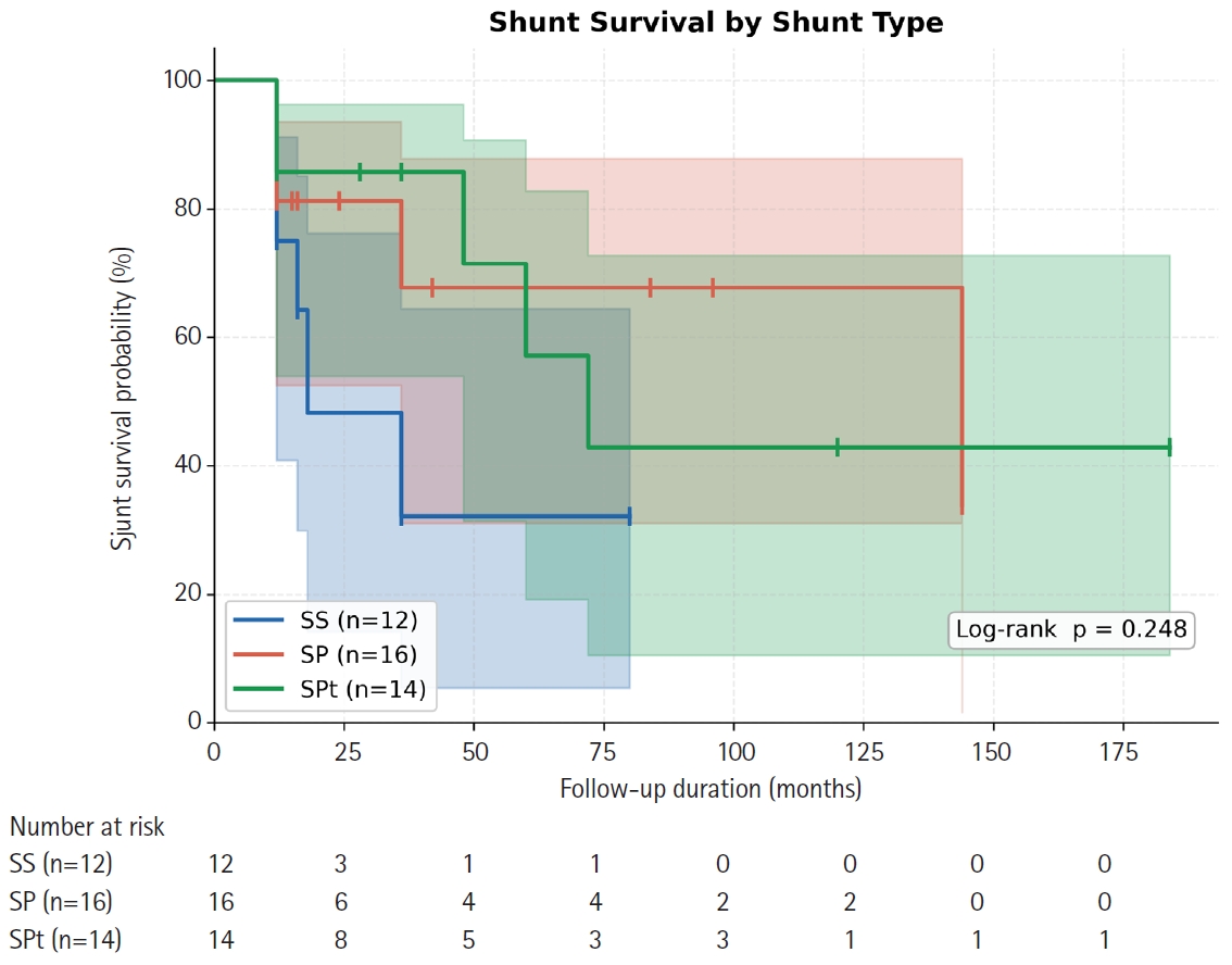
Purpose This study aimed to compare long-term shunt patency by shunt type (syringo-subarachnoid [SS], syringo-pleural [SP], syringo-peritoneal [SPt]) and disease etiology (post-traumatic, post-infectious, idiopathic) in patients surgically treated for syringomyelia, using Kaplan-Meier survival methodology.
Overview of Literature: Syringomyelia shunting carries a well-documented revision burden, but direct comparative survival data across shunt modalities and etiologic subgroups are scarce, particularly from East Asian centers.
Methods We retrospectively analyzed 42 patients (mean age, 47.6±11.8 years; 57.1% male) who underwent syringomyelia shunting at a tertiary neurosurgical center (January 2000–December 2020) with ≥12 months follow-up. Shunt type was classified as SS (n=12), SP (n=16), or SPt (n=14); etiology as post-traumatic (n=24), post-infectious (n=10), idiopathic (n=6), or hemorrhage/tumor-related (n=2). The primary endpoint was shunt revision surgery. Kaplan-Meier analysis, log-rank testing, and Cox proportional hazards regression were performed.
Results Over a median follow-up of 15 months (range, 12 to 184 months), 16 patients (38.1%) underwent shunt revision. Overall 12- and 24-month patency rates were 79.6% and 72.5%, respectively (median shunt survival 72 months). By shunt type, 12-month patency was 75.0% (SS), 81.3% (SP), and 85.7% (SPt); 24-month patency declined to 48.2% for SS while SP and SPt remained at 81.3% and 85.7% (log-rank p=0.248). Post-infectious syringomyelia showed the shortest median shunt survival (36 months) compared with post-traumatic (not reached) and idiopathic (not reached) groups (log-rank p=0.232). No independent predictor of shunt failure was identified on multivariate Cox regression.
Conclusion All three shunting techniques achieve approximately 80% one-year patency. SS may carry a higher long-term occlusion risk. Post-infectious syringomyelia requires closer postoperative surveillance. Larger prospective studies are needed to establish definitive shunt selection criteria.
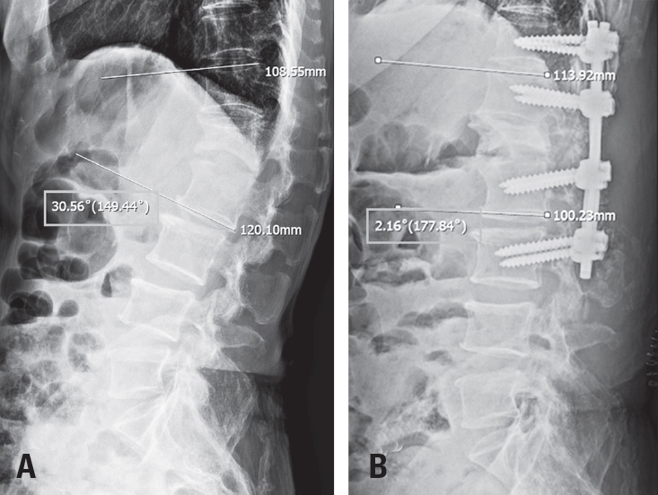
Tethered cord syndrome (TCS) is a condition in which the spinal cord becomes pathologically stretched due to various congenital or acquired etiologies, leading to progressive neurological symptoms. While surgical detethering is the gold standard for pediatric patients, adult-onset recurrent TCS presents a significant surgical challenge. Reoperation carries substantial risks—including spinal cord injury, cerebrospinal fluid leakage, and a high rate of retethering—often resulting in suboptimal long-term outcomes. Recently, spine-shortening osteotomy (SSO) has emerged as an alternative technique to reduce spinal cord tension without direct manipulation of the neural elements. Here, we report a case of recurrent adult TCS associated with a lipomyelomeningocele, which was exacerbated by post-traumatic kyphosis from an L1 compression fracture. The patient was successfully treated with SSO at the L1 level. This case highlights the utility of SSO as a safe and effective alternative to conventional revision detethering, particularly in complex cases involving spinal deformity.
Study Design A retrospective comparative study.
Purpose This study aimed to evaluate the reliability of the external auditory meatus (EAM) as a reference point for determining the cranial center in patients with sagittal spinal deformity. Specifically, we assessed the radiographic visibility and spatial relationship of the EAM relative to the midpoint of the nasion-inion line (MNI) and compared it with the sella turcica.
Overview of Literature: The cranial center is commonly defined as the midpoint of the nasion-inion line (MNI). The EAM has been widely used as an approximate surrogate of the MNI; however, its radiographic clarity and consistency are often limited. In contrast, the sella turcica is a singular anatomical structure that is more distinctly visualized on radiographic imaging.
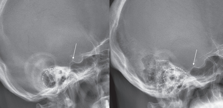
Methods A total of 71 patients who underwent corrective surgery for spinal deformity between 2021 and 2024 were included. Preoperative full-spine radiographs were analyzed to evaluate the spatial relationship between both the EAM and the sella turcica relative to the MNI. Radiographic visibility was classified into three categories: single-point and clear, duplex but distinguishable, and unclear.
Results The EAM was located, on average, 1.1 mm posterior and 17.1 mm inferior to the MNI, whereas the sella turcica was located 13.8 mm anterior and 2.0 mm superior to the MNI. Relative to the femoral head axis, angular analysis revealed that the EAM exhibited a mean posterior tilt of 0.2°, while the sella turcica showed a mean anterior tilt of 1.3°. The sella turcica was clearly identifiable in 84.5% of cases, whereas the EAM was clearly visualized in only 14.1%, with the majority being either duplicated or indistinct (p<0.001).
Conclusions Although the EAM is anatomically closer to the MNI, its radiographic visibility and consistency are inferior to those of the sella turcica, thereby reducing its reliability as a cranial reference point. Further studies are needed to determine whether the sella turcica can serve as a more reliable alternative reference.


 First
First Prev
Prev