Department of Neurosurgery, Inha University College of Medicine, Incheon, Korea
Corresponding author: Dalsung Ryu, M.D. Department of Neurosurgery, Inha University College of Medicine, 27 Inhang-ro, Jung-gu, Incheon 22332, Korea TEL: +82-32-890-3596, FAX: +82-32-890-2374, E-mail: dalsung.ryu@gmail.com
• Received: December 31, 2025 • Revised: March 13, 2026 • Accepted: April 7, 2026
This is an open access article distributed under the terms of the Creative Commons Attribution Non-Commercial License (https://creativecommons.org/licenses/by-nc/4.0/) which permits unrestricted non-commercial use, distribution, and reproduction in any medium, provided the original work is properly cited.
The incidence of compression fractures is increasing in aging populations. Differentiating pathological fracture types is complex and requires careful consideration during diagnosis. This case report describes the clinical course of a 54-year-old female patient presenting with progressive paraplegia after a back injury sustained while lifting a heavy object. Initial imaging revealed a burst fracture at T12 and severe spinal cord compression due to an epidural mass extending from T12 to L2. Clinical assessment raised suspicions of a hematologic malignancy or pathological fractures. Laminectomy and spinal fusion, along with mass removal, resulted in partial improvement in motor function and patient-reported pain levels. However, further evaluation and biopsy revealed chronic inflammation with fibrosis consistent with an unresolved hematoma. This case underscores the importance of a comprehensive differential diagnosis and multidisciplinary collaboration, integrating radiologic, surgical, and pathologic correlation, in the management of complex spinal pathologies.
Compression fractures of the spine can have various etiologies, including trauma, osteoporosis, and malignancy.1,2) The incidence of compression fractures is increasing with rapidly aging populations.1) Although high-energy impacts can lead to traumatic fractures, even minor trauma causes fractures in individuals with osteoporosis.1) Additionally, spinal column metastasis can lead to compression fractures.2) Previous studies have reported that approximately 5.5% of vertebral compression fractures (VCFs) are related to malignancy, as identified by routine biopsy, including both metastatic spinal lesions and primary spinal malignancy.3) Consequently, differentiating pathological fracture types is more complex than it initially appears and requires careful consideration during diagnosis.2,4,5) This case highlights the diagnostic challenges and treatment approach for a patient initially suspected of having a spinal malignancy.
Case Report
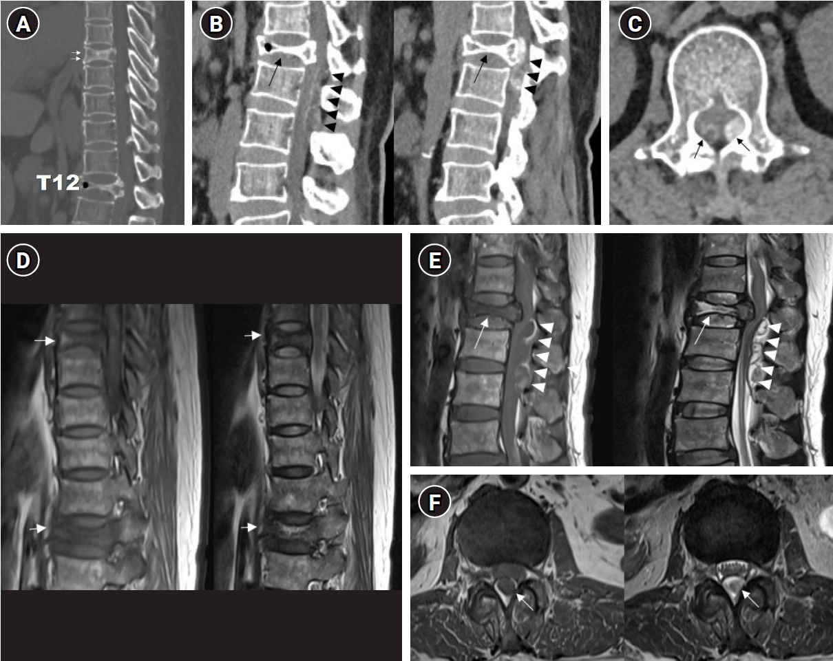
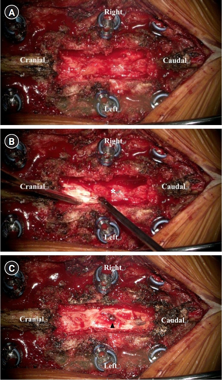
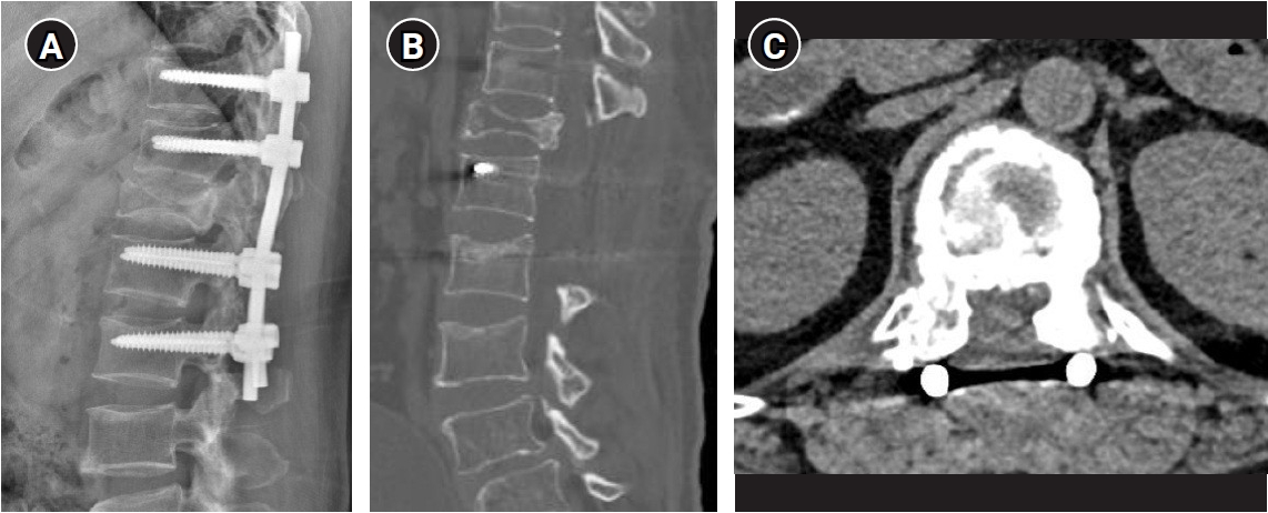
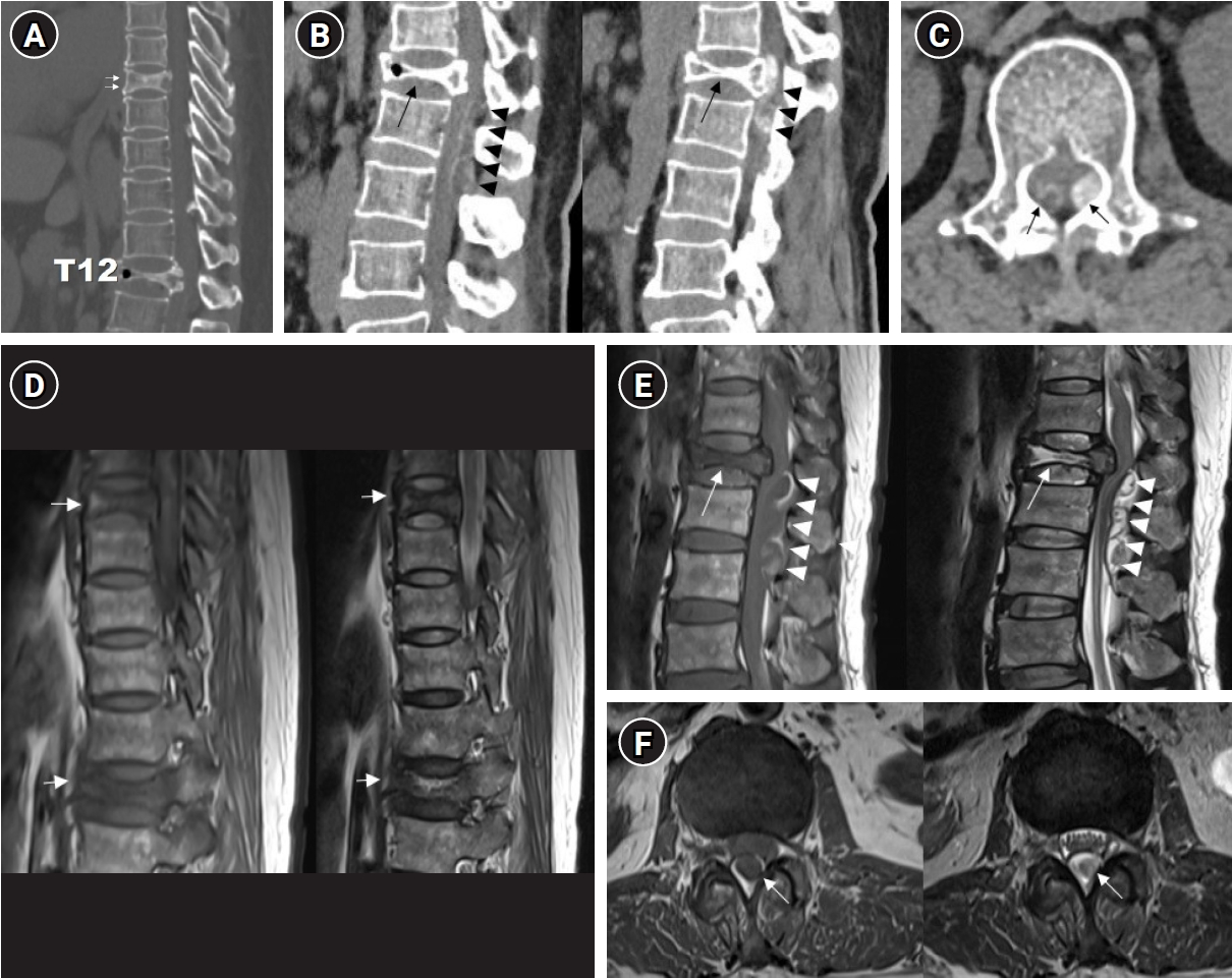
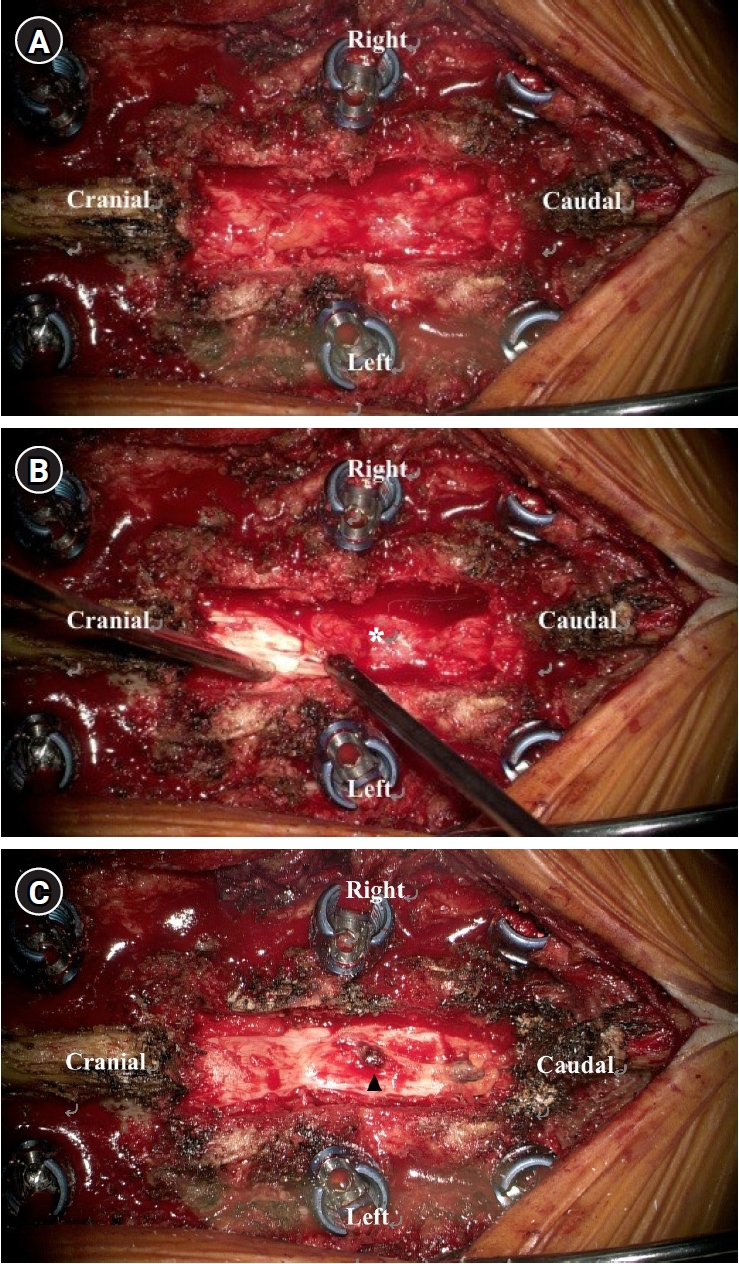
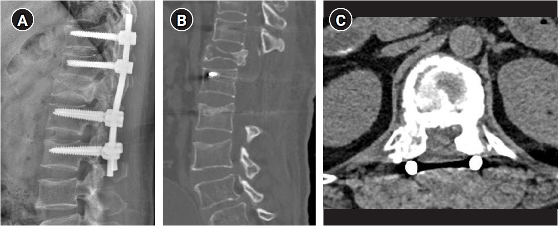
A 54-year-old woman presented to the emergency department with severe lower back pain, weakness in both legs, and difficulty ambulating after a fall. Her symptoms began a month earlier after lifting a heavy object, initially presenting with incomplete paraplegia that progressively worsened. Another fall injury 1 week prior to her visit exacerbated her symptoms. She had a history of complex psychiatric disorders, including depression and schizophrenia, but no other notable medical or neoplastic conditions. On physical examination, the patient’s right leg strength was graded as I, left leg strength as II across all joints, and low back pain was 7 on a numerical rating scale. She had voiding difficulty requiring urinary catheterization, whereas bowel function was intact. Thoracic and lumbar spine radiographs revealed a T12 burst fracture. Whole-spine computed tomography (CT) showed compression fractures at both T7 and T12 (Fig. 1A). The T12 lesion demonstrated features of a subacute or chronic burst fracture with retropulsion of the posterior vertebral body, and a mass in the posterior epidural space extending from T12 to L2 with severe central canal narrowing. Additionally, an ill-defined radiopacity was noted in the intradural and left anterior epidural spaces extending from T12 to L2. Although an iatrogenic cement leakage was initially suspected, the patient had no history of any spinal procedures. Among the differential diagnoses for epidural calcification - namely metastatic disease with epidural extension and arachnoiditis ossificans (AO)—the metastatic etiology appeared more likely (Fig. 1B, 1C).6) Initial magnetic resonance imaging (MRI) revealed compression fractures at T7 and T12 (Fig. 1D, 1E). The T12 fracture showed convex bowing of the posterior cortex with associated cord compression and an epidural mass. T1-weighted MRI revealed loss of normal marrow signal within the T12 vertebral body and an epidural mass causing cord compression. The mass appeared hyperintense, with a central area that was isointense to hypointense, resulting in central canal narrowing. T2-weighted imaging revealed that the T12 compression fracture was hyperintense and the epidural mass was heterogeneous but generally hyperintense with associated cord compression (Fig. 1E, 1F). Although contrast-enhanced MRI was recommended for further evaluation, it was not performed because the patient declined additional imaging studies. The patient’s laboratory results revealed thrombocytosis with a platelet count of 551 K/µL. Bone mineral density measurements indicated osteopenia, with a lumbar T-score of −1.6 and femoral neck T-score of −1.8. Further evaluations to rule out malignancy, including chest CT, abdominopelvic CT, positron emission tomography–CT, and tumor marker laboratory tests, showed no significant findings; however, a tissue biopsy was deemed necessary.3) The patient underwent a laminectomy, spinal fusion, and mass removal. The surgical procedures included total laminectomy of T12 and L1, subtotal laminectomy of the upper L2, pedicle screw fixation from T10 to L2, and posterolateral fusion. The epidural mass was carefully removed, and the fibrous tissue adherent to the dura was excised (Fig. 2). The surgery was completed after ensuring adequate decompression of the spinal cord. The pathological results of the laminectomized bone showed no evidence of malignancy. The epidural mass showed chronic inflammation with nodular fibrosis and bony detritus, indicating a chronic unresolved hematoma. On postoperative day 5, the patient’s right leg strength was graded I, while left leg strength improved to grade III for hip flexion and knee extension but decreased to grade I for ankle dorsiflexion. The patient had improved neurological status 1 month after discharge. Her left leg strength improved further to grade III for both ankles and great toe dorsiflexion, while the right leg remained at grade I. Postoperative imaging confirmed a well-decompressed state (Fig. 3).
This case report was conducted in accordance with the ethical standards of the institutional and national research committee and with the Helsinki Declaration. Institutional Review Board approval was not required for this case report. All patient information was fully anonymized, and no identifiable personal data were included in this manuscript.
Discussion
In this case, we initially suspected a pathologic spinal fracture with a malignant epidural tumor at admission; however, surgical histopathological examination revealed that the remaining mass was an unabsorbed hematoma. VCFs pose considerable diagnostic challenges, particularly when distinguishing benign and malignant osteoporotic fractures, which require vastly different management strategies and have different prognostic outcomes.1-5) In previous imaging studies, the characteristic imaging features observed on CT and MRI, including specific morphological changes, enhancement patterns, and MRI signal intensities, helped differentiate these fracture types.4,5) Benign osteoporotic VCFs typically show normal signals in the posterior elements, retropulsed bone fragments and are accompanied by additional benign fractures on MRI.4,5) These fractures also have preserved normal marrow signal with regular margins, and exhibit a linear horizontal hypointense T1/T2 band, fluid signs, and normal enhancement relative to adjacent vertebrae and after 3-month follow-up. On CT, benign fractures often display retropulsed bones, sharp fracture lines, puzzle signs, and an intravertebral vacuum.1,4,5) However, malignant VCFs show abnormal signals in the posterior elements, the presence of an epidural or paravertebral soft tissue mass, and an expanded posterior vertebral contour, often with metastasis to other vertebrae, on MRI.1,2,4,5) These fractures exhibit geographic replacement of the normal marrow signal, irregular margins, and increased enhancement relative to the adjacent vertebrae at 3-month follow-up.1,4,5) On CT, malignant fractures typically involve bone destruction and the presence of an epidural or focal paravertebral soft-tissue mass.1,2,4,5)
In this case, the initial imaging suggested a malignant process owing to the presence of T12 and T7 compression fractures, an epidural mass, and thrombocytosis. Whole-spine CT findings indicated calcifications and a posterior epidural mass extending from T12 to L1, which caused severe central canal narrowing. Additionally, an ill-defined radiopacity was noted in the intradural space and left anterior epidural space extending from T12 to the L2 level central canal. These findings initially suggested malignancy, given the abnormal posterior element signal, the presence of a mass with irregular margins, and bone destruction on CT.1,2,4,5) However, the biopsy results revealed chronic inflammation with fibrosis, consistent with an organized hematoma. Imaging findings suggested AO in the intradural space, which was not biopsied. This divergence from typical imaging features of malignancy highlights the complexities and potential pitfalls in VCF diagnosis. The patient presented with features common to both benign and malignant VCFs, including severe back pain and paraplegia. The presence of calcification within the epidural mass, which is not typically observed in benign osteoporotic fractures, initially suggested a malignancy. Conversely, the organized hematoma and chronic inflammation observed on biopsy indicate a benign process secondary to trauma. Subacute hematomas can be difficult to detect by CT imaging, and gradually appear isodense over time.7,8) However, MRI can provide a clearer view, revealing the form of the hematoma and enabling a precise assessment of the affected cord levels.7,8) MRI results can also indicate bleeding duration; acute hematomas typically appear hypointense on T1-weighted images and hyperintense on T2-weighted images.7-9) As the hematoma progresses to the early subacute stage, it becomes hyperintense on T1-weighted MRI and hypointense on T2-weighted MRI.7-9) Over time, the hematoma becomes increasingly hyperintense on T1-weighted sequences and hypointense on T2-weighted sequences, often exhibiting a mosaic pattern.7-9)
In the present case, the posterior epidural mass was identified as an organized hematoma consistent with traumatic spinal epidural hematoma (TSEH). TSEH involves bleeding primarily from the posterior epidural venous plexus, which is susceptible to rupture due to its lack of valves and proximity to pressure fluctuations.7-10) This network is particularly vulnerable at cervicothoracic and thoracolumbar junctions.7,9,11,12) Clinical cases have linked TSEH to activities such as heavy lifting, straining, and hypertension.7-9,11,12) However, some researchers, such as Beatty and Winston13), have suggested that arterial sources may be responsible for the bleeding, arguing that venous hemorrhage would not generate sufficient pressure to compress the spinal cord. Both TSEH and non-TSEH can present with acute pain followed by rapidly progressive neurologic deficits, and timely decompression is critical for favorable outcomes.7,8,10,11,14)
The presence of AO added further complexity to this case, owing to its rarity and difficulty in accurately diagnosing it. AO involves ossification within the subarachnoid space, potentially leading to the obstruction of cerebrospinal fluid flow and spinal cord compression.6) Non-contrast CT is particularly effective in demonstrating the extent of ossification and complements MRI findings.6) Although the diagnosis of AO was based on imaging rather than biopsy, the presence of ossified lesions complicated the clinical picture.
This case highlights the importance of integrating radiologic, surgical, and pathologic findings in diagnostic evaluation. Although advanced imaging techniques provide valuable insights, they must be interpreted cautiously and correlated with clinical and histopathological findings to avoid misdiagnosis.1-5) Distinguishing benign and malignant VCFs remains challenging and requires careful consideration of all diagnostic modalities.1-5) Furthermore, maintaining a high index of suspicion and utilizing a multidisciplinary approach is essential to achieve an accurate diagnosis and appropriate management.
In conclusion, this case report underscores the importance of distinguishing an unresolved epidural hematoma from a malignancy via surgical resection. Suspicion of malignancy considerably influences the decision-making processes of both the medical team and the patient's caregivers, impacting the overall treatment plan. Although mass formation with compression fractures is rare, it can occur as a result of trauma. Hence, careful attention must be paid to the diagnosis to avoid misinterpretation and ensure appropriate management.
The authors have no conflicts of interest to declare.
Funding
None.
Acknowledgments
None.
Fig. 1.
Preoperative imaging. (A) Whole-spine computed tomography (CT) sagittal image demonstrating compression fracture at T7 (arrows) and T12. (B) Whole-spine CT sagittal image showing a compression fracture at T12 (arrows) and an epidural mass (arrowheads). (C) Whole-spine CT axial image at the T12 level. The mass (arrows) consisted of a hyperdense component on the left and a hyperdense capsule with an isodense to slightly hypodense interior on the right. (D) The magnetic resonance imaging (MRI) T1, T2-weighted sagittal sequences demonstrating compression fractures at T7 and T12 (arrows). (E) The MRI T1, T2-weighted sagittal sequences demonstrating T12 compression fractures (arrows) and an epidural mass (arrowheads). (F) The MRI T1, T2-weighted axial sequences at the L1 level showing an epidural mass (arrows).
Fig. 2.
Intraoperative images. (A) Post-laminectomy image showing the decompressed spinal canal with fibrous tissue adherent to the dura. (B) Image showing the removal of fibrous tissue (asterisk) adherent to the dura mater, which appears elongated and torn. (C) The image shows the spinal region after extending the laminectomy to the upper L2 level. Complete decompression was achieved with the removal of all fibrous tissue. Areas of old hemorrhage (arrowhead) are visible, appearing as clumped regions.
Fig. 3.
Postoperative imaging. (A) Postoperative X-ray indicating spinal fusion from T10 to L2, demonstrating successful instrumentation and alignment post-surgery. (B) Postoperative computed tomography (CT) sagittal scan showing a well-decompressed state at the T12, L1, and upper L2 levels. The previously noted calcified lesions are no longer visible. (C) Postoperative CT axial scan at the T12 level, confirming the absence of the calcified lesion on the central canal and effective decompressive laminectomy.
References
1. Gutierrez-Gonzalez R, Royuela A, Zamarron A. Survival following vertebral compression fractures in population over 65 years old. Aging Clin Exp Res 2023;35:1609-17.
2. Taylor TN, Bridges CS, Pupa LE, Morrow BA, Smith BG, Montgomery NI. Retrospective review of 181 patients with pathologic vertebral compression fractures. J Pediatr Soc North Am 2023;5:697.
3. Mukherjee S, Thakur B, Bhagawati D, et al. Utility of routine biopsy at vertebroplasty in the management of vertebral compression fractures: a tertiary center experience. J Neurosurg Spine 2014;21:687-97.
4. Mauch JT, Carr CM, Cloft H, Diehn FE. Review of the imaging features of benign osteoporotic and malignant vertebral compression fractures. AJNR Am J Neuroradiol 2018;39:1584-92.
5. Abdel-Wanis ME, Solyman MT, Hasan NM. Sensitivity, specificity and accuracy of magnetic resonance imaging for differentiating vertebral compression fractures caused by malignancy, osteoporosis, and infections. J Orthop Surg (Hong Kong) 2011;19:145-50.
6. Nagashima Y, Nishimura Y, Ito H, Nishii T, Oyama T, Saito R. Diagnosis and treatment strategies for arachnoiditis ossificans following subarachnoid hemorrhage: a case report. NMC Case Rep J 2022;9:295-9.
8. Fedor M, Kim ES, Ding K, Muizelaar JP, Kim KD. Spontaneous spinal epidural hematoma: a retrospective study on prognostic factors and review of the literature. Korean J Spine 2011;8:272-82.
12. Alrazooqi MK, Skikic E, Iqbal SS, Sulaiman L, Muhammed Noori OQ. Traumatic spinal epidural hematoma with significant neurologic findings: a case report. Cureus 2023;15:e38869.
14. Kahraman MA, Senturk S. The necessity of extensive decompression for spinal epidural hematoma: a case report and literature review. Cureus 2023;15:e44192.
Unresolved Chronic Epidural Hematoma with Burst Fracture Mimicking Malignancy in a Patient with Paraplegia: A Case Report
Fig. 1. Preoperative imaging. (A) Whole-spine computed tomography (CT) sagittal image demonstrating compression fracture at T7 (arrows) and T12. (B) Whole-spine CT sagittal image showing a compression fracture at T12 (arrows) and an epidural mass (arrowheads). (C) Whole-spine CT axial image at the T12 level. The mass (arrows) consisted of a hyperdense component on the left and a hyperdense capsule with an isodense to slightly hypodense interior on the right. (D) The magnetic resonance imaging (MRI) T1, T2-weighted sagittal sequences demonstrating compression fractures at T7 and T12 (arrows). (E) The MRI T1, T2-weighted sagittal sequences demonstrating T12 compression fractures (arrows) and an epidural mass (arrowheads). (F) The MRI T1, T2-weighted axial sequences at the L1 level showing an epidural mass (arrows).
Fig. 2. Intraoperative images. (A) Post-laminectomy image showing the decompressed spinal canal with fibrous tissue adherent to the dura. (B) Image showing the removal of fibrous tissue (asterisk) adherent to the dura mater, which appears elongated and torn. (C) The image shows the spinal region after extending the laminectomy to the upper L2 level. Complete decompression was achieved with the removal of all fibrous tissue. Areas of old hemorrhage (arrowhead) are visible, appearing as clumped regions.
Fig. 3. Postoperative imaging. (A) Postoperative X-ray indicating spinal fusion from T10 to L2, demonstrating successful instrumentation and alignment post-surgery. (B) Postoperative computed tomography (CT) sagittal scan showing a well-decompressed state at the T12, L1, and upper L2 levels. The previously noted calcified lesions are no longer visible. (C) Postoperative CT axial scan at the T12 level, confirming the absence of the calcified lesion on the central canal and effective decompressive laminectomy.
Fig. 1.
Fig. 2.
Fig. 3.
Unresolved Chronic Epidural Hematoma with Burst Fracture Mimicking Malignancy in a Patient with Paraplegia: A Case Report